Immediate Effects of Appling Resistance in the Bridge Exercise on Muscle Activity in the Trunk and Lower Extremities
© 2023 by the Korean Physical Therapy Science
Abstract
The bridge exercise prevents repeated damage to the tissues around the spine by reducing stimulus transmission to the ligaments and joint capsules, thereby alleviating back pain. It also contributes to strengthening the muscles of the lower extremities.
A Single Subject experience design.
This study was conducted in 28 healthy adults in their 20s to 30s and conducted at St. Mary's Hospital in C City from May to July 2021. Four types of bridge exercise were performed in this study: the normal bridge exercise and bridge exercises with 0.5%, 1%, or 1.5% body weight resistance applied on the pelvis through manual resistance during the bridge exercise and to determine the effect of resistance applied in the bridge exercise on the activation of the trunk and lower extremities muscles.
This study showed that the muscle activity of the trunk and lower extremities improved significantly in response to stronger resistance when manual resistance equivalent to 0.5%, 1%, or 1.5% of body weight was applied during the bridge exercise compared to when the normal bridge exercise was performed.
This study is that manual resistance can be applied as an effective method of bridge exercise since muscle activity in the trunk and lower extremities increases when manual resistance causing isometric contraction is applied.
Keywords:
bridge exercise, electromyography, muscle activity, manual resistanceⅠ. Introduction
The bridge exercise performed with one’s knees bent in a comfortable position while lying on a back, after which the hips are raised (Jung et al, 2017). Bridge exercise is a foundational exercise in most physical activities or sports that emphasize the body’s stability, as it can enhance physical stability through simultaneous contraction of the muscles around the trunk. The bridge exercises relieve back pain by reducing the transmission of impulses to ligaments and joint capsules, preventing repetitive damage to tissues around the spine (Stevens et al, 2007). Furthermore, it helps to absorb external forces exerted on the back, leading to coordination and complementation between the muscles around the back (Jeon, 2010,Oh, 2022). The bridge exercise is a closed kinematic chain exercise that increases spinal and pelvic stability by activating the muscles connected to the lumbo-pelvic area. The exercise can be done through movements necessary for activities of daily living and partial weight-bearing necessary for ambulation (O'Sullivan et al, 2019).
The bridge exercise is applied to increase the strength of the gluteus maximums and hamstring in an effort to promote stabilization of the back and trunk (Kisner et al, 2017), and it uses various synergic muscles and stabilizer muscles such as the erector spinae, rectus abdominis, external oblique, and internal oblique (Moon, 2017). Accordingly, the importance of body stabilization and the necessity of the bridge exercise, which requires activating the muscles of the trunk, have been emphasized in rehabilitation, (Lehman et al, 2005). This exercise involves selectively applying muscle activation of the lower extremities while maintaining stabilization of the trunk (Shumway-Cook et al, 2001), and the muscles that are activated may vary according to the type of the intervention. Previous studies have investigated isometric contraction in the adductor muscle group of the hip joint, changes of the knee joint angle, modified bridge exercises, and changes in the support plane (Kim et al, 2010; Kim and Hwang, 2013; Lee et al, 2014; Lee et al, 2010; Han et al, 2015). The bridge exercise has also been done using various instruments such as Swiss balls, balance pads, and slings to enhance the core muscles (Kim et al, 2018; Kim et al, 2012; Kim et al, 2016; Kim et al, 2009; Song et al, 2011).
A recent study also investigated the activation of abdominal muscles during the bridge exercise through weights (loads) or manual correction (Moraes et al, 2009; Kim et al, 2014). According to a study on the effect of different types of foot support on the muscle activity of the trunk and leg muscles (Kim et al, 2016), sole support led to higher activity of the erector spinae, gluteus maximus, and semitendinosus muscles than the other muscles, toe support increased the activity of the semitendinosus and soleus muscles, and heel support influenced the activity of the soleus muscle. In addition, compared to the normal bridge exercise, the bridge exercise with the soles pressing on the ground without changing the angle of the ankles led to higher muscle activity in the erector spinae, gluteus maximus, semitendinosus, peroneal, rectus femoris, and rectus abdominis muscles and showed statistically significant differences (Jung et al, 2017). With the method of foot support and pressing the floor with the soles, a study investigated the activity of the abdominal muscles when using weights (loads) or manual correction during the bridge exercise and found that the group that adjusted their posture following the hand of the therapist had a significantly higher gluteus maximus/hamstring muscle activity ratio than the control group (Moraes et al, 2009; Kim et al, 2014). Moreover, Park et al. (2013) reported that when participants put their hand on the anterior superior iliac spine and corrected the posture of their pelvis while they actively raised their legs in a lying position, pelvic rotation was reduced and the activity of the abdominal muscles increased compared to participants who used a belt or did not adjust their posture.
A study on the activity of the trunk and leg muscles when various weights were applied to modified bridge exercises reported that when, a weight equivalent to 0.5% of body weight was loaded, the activity of the vastus medialis muscle and gluteus medius increased (Lee, 2019). As in the above study, performing the exercise with a weight applied to a specific body part can minimize boredom and enhance the motivation of the exerciser (Kim, 2005), and adding weights to the body weight and on body segments can increase the physical exercise performed by each body segment (Jun, 1994). A previous study reported that weight-bearing exercises can induce effects in smaller (in terms of speed or the range of motion) types of movement and improve the effects in people who have limited physical movement when they move within their available range of motion (Yang, 1996). Although there are various ways of applying weight loads, Cho et al. (2016) reported that manual resistance is advantageous in that it can reduce back pain, control the intensity of the exercise according to patients’ condition, draw forth their exercise capacity, and allow one to perceive the muscular strength of patients while they are exercising (Cho et al, 2016).
The bridge exercise is currently widely used in clinical practice for trunk stabilization, and the intensity of the training is controlled by applying manual resistance to the pelvic area. However, in previous studies, manual resistance and weight loads were mainly applied to the arms or legs (Lee 2017, Park et al 2021), and there are insufficient studies where manual resistance and weight loads were applied for trunk stabilization during the bridge exercise. Moreover, since prior studies have stated that the method where a therapist applies manual resistance is widely performed in clinical practice, it can be inferred that better results will be observed if it is used in conjunction with diverse methods of exercise therapy (Cho et al 2016). Therefore, this study aimed to analyze the muscle activity of the trunk and lower extremities when various weights (loads) were applied on the pelvis through manual resistance during the bridge exercise and to determine the effect of resistance applied in the bridge exercise on the activation of the trunk and lower extremities muscles. Another purpose of this study was to increase the effect of the bridge exercise and provide the basic data necessary for creating spinal stabilization training programs.
Ⅱ. Methods
This study was conducted in 28 healthy adults in their 20s to 30s, and the participants in this study received a sufficient explanation of the purpose and methods of this study before the experiment, filled out an informed consent form indicating their consent to the purpose of the study, and expressed willingness to participate in the study before joining the experiment. The inclusion criteria were people who did not have back pain, musculoskeletal disease, a surgical history, a limited range of motion of the back and lower extremity joints, or a loss of muscle strength. Those who had neurological conditions, cardiopulmonary diseases, or congenital deformities of the arms and legs were excluded. Table 1 presents the general characteristics of the participants.
After being approved by the IRB of the Korea National University of Transportation (KNUT IRB 2021-27), this study was conducted at St. Mary’s Hospital in C City from May to July 2021. Four types of the bridge exercise were performed in this study: the normal bridge exercise and bridge exercises with 0.5%, 1%, or 1.5% body weight resistance applied. In the normal bridge exercise, one fixes the knees at 60° in a supine position, raises the hip joint to 0°, and holds for 5 seconds at the verbal cue of the therapist. In the bridge exercises with resistance applied, 0.5%, 1%, or 1.5% of body weight was applied by the therapist through manual resistance during the bridge exercise. When performing each type of the bridge exercise, the activity of the bilateral erector spinae, rectus abdominis, gluteus medius, hamstring, and tibialis anterior muscles was measured using a wireless surface electromyography system.
All participants were given a 10-minute break between measurements to prevent muscle fatigue and injury when performing the four types of the bridge exercises, and research assistants stood by during the experiment to prevent dangerous situations.
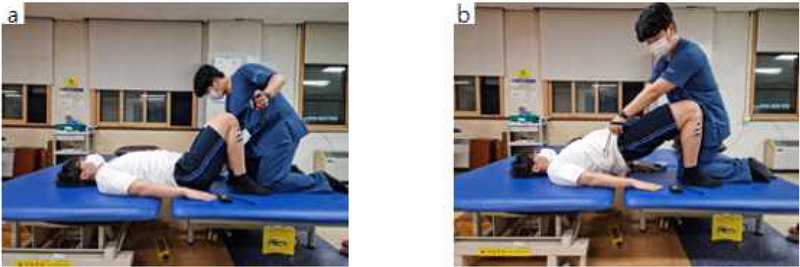
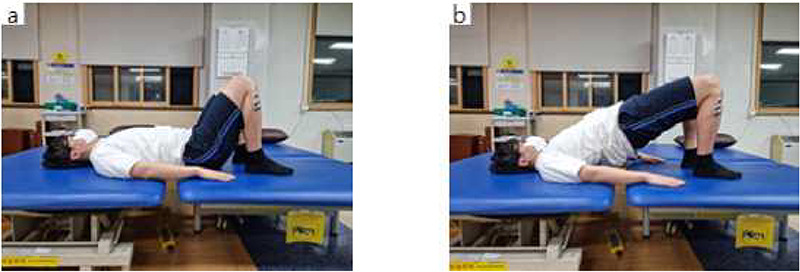
The setup for the normal bridge exercise is to lie down with the knees raised at 60°, both feet spread at shoulder width parallel to each other and touching the floor, both arms straight on the supporting plane, and the head and eyes fixed on the ceiling. When the participant is ready to perform the bridge exercise, he/she raises the hips to the verbal cue “raise your hips” until the hip joint is at 0° and holds it for 5 seconds. Resistance is not applied at this stage (Figure 1). In the variations of the bridge exercise with resistance applied, the participant raises the hips from the setup for the normal bridge exercise in response to the verbal cue “raise your hips” until the hip joint is at 0°, holds it for 5 seconds, and the therapist applies resistance to the both sides of the participant’s pelvis while wearing digital myometers on both hands (Figure 2).

isometric contraction bridge exercise : (a) Before the isometric contraction bridge exercise (0.5%, 1%, 1.5%/BW) (b) During the isometric contraction bridge exercise (0.5%, 1%, 1.5%/BW)
After the therapist applies resistance to the anterior superior iliac spine area while the participant maintains the bridge exercise posture, he/she applies manual resistance using both hands and measures the isotonic contraction. Manual resistance equivalent to 0.5%, 1%, or 1.5% of body weight was displayed visually on a digital myometer(Jtech Commander Echo, JTECH Medical, USA) (Figure 3).
Measurement
Muscle activation of both trunk and lower limbs was measured using wireless surface electromyography (sEMG) (freeEMG1000, BTS Bioengineering, Milano, Italy). The sample rate and bandpass filter were set to 1,000 and 20–500 Hz, respectively. Wireless sEMG electrodes were attached to both lower limb muscles (erector spinae, rectus abdominis, gluteaus medius, hamstring, tibialis anterior) using adhesive electrodes. Ten wireless adhesive electrodes were attached to the five major muscle groups of both trunk and lower limbs based on the SENIAM (Surface EMG for Non-Invasive Assessment of Muscles) guideline20. Muscle activation data were obtained using an EMG Analyzer v2.9.37.0 (BTS Bioengineering, Milano, Italy). To remove artifacts and high-frequency noise, the obtained sEMG raw data were bandpass filtered (20–500 Hz). All values were set to Maximal Voluntary Contraction (MVC) and expressed as %MVC to normalize the sEMG signal(Cram et al, 1998). For analysis, 6-11 seconds were analyzed except for 5 seconds in the preparation posture during the 11-second measurement.
Statistical analysis
Data analysis was performed using SPSS version 21.0. (IBM Corp, Armonk, NY, USA). Descriptive statistics were used to analyze the general characteristics and baseline clinical assessments of the participants. The Kolmogorov-smirnov test was used to confirm that all outcome variables were normally distributed. For dependent variable measures, repeated measures analysis of variance (post hoc analysis used Bonferroni correction) was used to compare changes in the muscle activation of both trunk and lower limbs and were conducted to compare the muscle activity of the trunk and lower extremities according to general bridge exercise and bridge exercise with resistance (0.5%, 1%, 1.5% of body weight) applied. A significance level of 0.05 was used for all tests
Ⅲ. Results
In total, four bridge exercises were applied to 28 subjects who participated in this study. In this study, when bridge exercise with resistance of 0.5%, 1%, and 1.5% were applied, the muscle activity of the erector spinae muscle, rectus abdominis muscle, gluteus medius, hamstring and anterior tibialis muscle increased as the resistance increased and It can be seen that there was a significant improvement in comparison between groups (p=.000)(Table 2).
Ⅳ. Discussion
Strength training in recent clinical practice for musculoskeletal patients has focused on the accurate recruitment of weakened muscles and enhancement of motor control ability. Through various methods, such as posture correction or manual resistance by the therapist, for the recruitment of accurate muscles, compensation movements are minimized at a level of difficulty appropriate for the patient’s capacity in order to facilitate effective rehabilitation. Accordingly, this study aimed to determine how the application of manual resistance during the bridge exercise, which is commonly used in trunk stabilization training, would impact muscle activity in the trunk and lower extremities. In this study, muscle activity was measured during the normal bridge exercise and variations of the bridge exercise with 0.5%, 1%, or 1.5% resistance applied. The bridge exercise is a closed kinematic chain weight-bearing exercise widely used in clinical practice to increase the strength of lower extremities (Kisner et al, 2007), stabilize the trunk, and increase the strength of the glutes and legs.
This study showed that when manual resistance equivalent to 0.5% of body weight was applied, the muscle activity of both erector spinae muscles substantially improved as the manual resistance increased. Although the muscle activity increased as a result of resistance equivalent to 1% and 1.5% of body weight, there was no significant difference (p>.05). The activity of the erector spinae is thought to have increased because the bridge exercise stabilizes the spine and can activate muscles sufficiently with resistance equivalent to just 0.5% of body weight while one holds the posture with the resistance applied. Among the local muscles that are involved in trunk flexion during the bridge exercise, the erector spinae accounts for a large proportion of trunk flexion (Lee et al 2015). Muscle activity increased during the bridge exercise (Neumann, 2010) as a result of contraction of the gluteus maximus to the end of the range of motion of the hip joint and the movement done to raise the hips to the target level or bear the weight resistance.
The rectus abdominis muscle activity improved significantly when manual resistance equivalent to 0.5%, 1%, or 1.5% of body weight was applied compared to the normal bridge exercise. It is thought that the improved activity of the rectus abdominis as the load increased progressively is in accordance with the overload principle, since the rectus abdominis plays a role in maintaining balance against gravity or external loads exerted on the body, as well as the stability of the trunk. A study on muscle loading and spinal stabilization in healthy adults during lumbar stabilization exercises by Kavcic et al. (2004) reported that the bridge exercise was highly correlated with the muscle activity of the rectus abdominis. Although only the superficial muscle groups are activated by the normal bridge exercise, applying manual resistance as in this study can activate the deep muscles as well. In addition, since raising the hips during the bridge exercise increases the intra-abdominal pressure and provides dynamic stabilization for the rotation and translational stress of the back, contraction of the rectus abdominis provides optimal neuromuscular efficiency (Hodges et al, 1999).
The muscle activity of the gluteus medius increased significantly when manual resistance equivalent to 0.5%, 1%, or 1.5% of body weight was applied compared to the normal bridge exercise, which agrees with the results of previous studies. A study by Lee (2017) found significant differences between 0% to 0.5% and 0% to 1% of body weight when the bridge exercise was performed using weight loads, and 0.5% was reported to be an appropriate weight for selective contraction of the gluteus medius. It is believed that contraction of the gluteus medius stabilized the muscle between the hip joint and the spine similarly to the gluteus maximus during the bridge exercise, and a high level of activity of the gluteus medius may be necessary in various environments that are more dynamic than the normal bridge exercise and where the exerciser has to bear resistance. These weight-bearing exercises are beneficial in that the activity of the muscles can be increased (Bolgla et al, 2005). The gluteus medius is a very important muscle for therapeutic rehabilitation exercises of the hip joint in actual clinical practice, and to enhance the strength of the gluteus medius and improve its muscle activity, exercises should be performed through appropriate loading, such as the progressive loading protocol. The important role of the gluteus medius and the necessity of strengthening the muscle are emphasized in actual clinical practice to stabilize the pelvis and minimize knee misalignment during weight-bearing exercises (Ayotte et al, 2007). Since it is important to improve the strength of the gluteus medius, the therapist should help the patient maintain a posture that activates the gluteus medius and provide appropriate loads when applying exercises to a patient with hip joint problems. As manual resistance was applied on the pelvis in this study, the actions for bearing isometric resistance stabilized the sacroiliac joint, placed pressure on the joint surface to enhance the form closure, and enhanced the selective increase of muscle activity. Appropriate muscle activity of the hip joint muscles and pelvic area contributes to pelvic stabilization and influences the muscle activity of the muscles around the pelvis and back. A previous study reported that external support of the pelvis selectively increased the activity of the gluteus medius (Lee et al, 2011), and in another prior study, when isometric contraction was applied for hip abduction during the bridge exercise, the activity of the gluteus maximus and gluteus medius increased by 10.88% and 4.75%, respectively. Isometric resistance applied during the bridge exercise led to significant improvements in the gluteus medius, which correlates with the result of previous studies (Choi et al, 2015).
The muscle activity of the hamstring and tibialis anterior muscle improved significantly when manual resistance equivalent to 0.5%, 1%, or 1.5% of body weight was applied compared to the normal bridge exercise. The bridge exercise is widely used as an intervention method for improving trunk stability and strength by enhancing the coordination of superficial muscles (e.g., the erector spinae and hamstring) and deep muscles (e.g., the multifidus muscles and gluteus maximus) (O’Sullivan et al, 2007). It has been reported that the bridge exercise develops the coordination patterns of muscles by activating local and deep muscles at an appropriate ratio (Stevens et al., 2006), enhances neuromuscular control for trunk flexion and extension, and improves the functional stability of the trunk and back area by strengthening the pelvis and leg muscles (Kisner et al, 2007). Strengthening the lower extremity muscles during the bridge exercise caused significant improvements in the muscle activity of the hamstring and tibialis anterior muscle when manual resistance was applied.
Unlike previous studies, muscle activity was measured bilaterally in this study, and the muscle activities on both sides were similar due to the direct and symmetrical application of manual resistance by the therapist on the pelvis. When applying the bridge exercise, it is important to maintain proper alignment and a neutral position of the pelvis and spine through the simultaneous contraction of the deep spinal muscles, and if the correct movement is not performed, it may result in excessive forward bending of the back or asymmetrical trunk muscles and leg muscles (Stevens et al., 2006). Therefore, since it is of utmost importance to control the level of difficulty of the exercise and apply the exercise to patients carefully according to their level, it is considered that fixing the patient’s pelvis while applying manual resistance to it helped maintain a neutral posture. Moreover, the reason why the muscle activity of the hamstring and tibialis anterior muscle was relatively low is believed to be because the agonistic muscles during the bridge exercise are the gluteus medius and erector spinae, which extend the hip joint. In actual clinical practice, the activation patterns of the hamstring are very important. The data of this study on activation of the hamstring during the bridge exercise can be very beneficial for preventing hamstring injuries in athletes or normal people, and low manual resistance (equivalent to 0.5% of body weight) enable sufficient activation of the hamstring of patients in early rehabilitation after surgery, and muscle activity increases when the posture is held against resistance. In addition, the bridge exercise has the advantages that it can be applied (with or without partial weight loads) to people with pelvis, knee, and ankle instability, as well as patients who need to strengthen the hamstring group during rehabilitation exercises related to injuries of the anterior cruciate ligament in the lower extremities. Since the gluteus medius, hamstring, and tibialis anterior muscle create synergy in raising the pelvis during the bridge exercise (Choi et al, 2015), change in the activity of a muscle can be related to increased activity of the elevating muscles (Jonkers et al, 2003). Although the bridge exercise with manual resistance was performed by uninjured adults in this study, it is believed that this exercise can increase the physical exercise conducted by each body segment by increasing the body weight or body segment weight during rehabilitation training for patients with musculoskeletal injuries or neurological injuries.
This method can also minimize the boredom experienced by patients during the exercise, and raise their motivation by applying specific weight loads on the body. This study has the limitation that it the findings are difficult to generalize since the number of the participants was relatively low and it was conducted among healthy participants. Also, since there are more male subjects than female subjects, it may have affected the increase in muscle strength. Studies that take this limitation into account will be needed in the future. Nevertheless, the results of this study showed that the bridge exercise, which caused isometric contraction, led to significant improvements in muscle activity in the trunk and lower extremities. These findings will provide the basic data necessary to organize spinal stabilization programs in clinical practice.
Ⅴ. Conclusion
This study was conducted in 28 healthy adults to analyze the effect of the bridge exercise, which causes isometric contractions, on muscle activity in the trunk and lower extremities. The muscle activity of the trunk and lower extremities improved significantly in response to stronger resistance when manual resistance equivalent to 0.5%, 1%, or 1.5% of body weight was applied during the bridge exercise compared to when the normal bridge exercise was performed. In addition, muscle activity increased significantly at the minimum resistance, equivalent to 0.5% of body weight. Manual resistance can be applied as an effective method of the bridge exercise, since the muscle activity in the trunk and lower extremities increases when manual resistance causing isometric contraction is applied.
References
-
Ayotte NW, Stetts DM., Keenan, G. Greenway, EH 2007 Electromyographical analysis of selected lower extremity muscles during 5 unilateral weight-bearing exercises. Journal of Orthopaedic & Sports Physical Therapy 37(2): 48-55.
[https://doi.org/10.2519/jospt.2007.2354]

-
Bolgla LA, Uhl TL 2005 Electromyographic analysis of hip rehabilitation exercises in a group of healthy subjects. Journal of Orthopaedic & Sports Physical Therapy 35(8): 487-494.
[https://doi.org/10.2519/jospt.2005.35.8.487]

- Cho GT, Lee WJ, Gwon YB 2016 The Effect of Manual Resistive Exercise Therapy on Chronic Low Back Pain Patients. Joulinal of Sport Science 2(1): 39-49.
- Cram JR, Kasman GS, Holtz J 1998 Introduction to surface electromyography. Aspen publishers.
-
Gku Bin Oh et al. Analysis of trunk and lower extremity muscle activity according to the compensation of arm during bridge exercise. J of Kor Phys Ther 2022;29(3):12-20.
[https://doi.org/10.26862/jkpts.2022.09.29.3.12]

-
Hodges PW, Richardson CA 1999 Transversus abdominis and the superficial abdominal muscles are controlled independently in a postural task. Neuroscience letters 265(2): 91-94.
[https://doi.org/10.1016/S0304-3940(99)00216-5]

- Yang JS 1996 Physiological responses to walking exercise with added weight at different locations in normal subjects. International journal of human movement science 35(1): 130-143.
- Jeon HY 2010 The effects of a bridging exercise on body shape changes and foot pressure distribution. Daegu University. Korea.
-
Jonkers I, Stewart C, Spaepen A 2003 The complementary role of the plantarflexors, hamstrings and gluteus maximus in the control of stance limb stability during gait. Gait & posture 17(3): 264-272.
[https://doi.org/10.1016/S0966-6362(02)00102-9]

- Jun JK 1994 Cardiorespiratory responses to walking with trunk weights. The Korean Journal of Physical Education 33(1): 331.
-
Jung MK, Kang KH 2017 The Effect of Bridge Exercise with Pressing Ground on Activation of Lower Body Muscle and Trunk Muscle. Journal of Sport and Leisure Studies 68: 593-600.
[https://doi.org/10.51979/KSSLS.2017.05.68.593]

-
Kavcic N, Grenier S, McGill SM 2004 Quantifying tissue loads and spine stability while performing commonly prescribed low back stabilization exercises. Spine 29(20): 2319-2329.
[https://doi.org/10.1097/01.brs.0000142222.62203.67]

-
Kim DH, Choi GR 2016 Effect of Bridge Exercise with Different Foot Stances on Trunk and Lower Limbs Muscle Activity.Journal of Sport and Leisure Studies 66:715-722.
[https://doi.org/10.51979/KSSLS.2016.11.66.715]

-
Kim DH, Kim TH 2018 The Effects of Sling and Resistance Exercises on Muscle Activity and Pelvic Rotation Angle During Active Straight Leg Raises and Pain in Patients with Chronic Low Back Pain. J Korean Soc Phys Med 13(4): 113-121.
[https://doi.org/10.13066/kspm.2018.13.4.113]

-
Kim JW, Hwang BJ 2013 Comparison of Muscle Activity of Lower Limbs in Bridging Exercise according to Thigh Adduction-Abduction and Tibia Internal-External Rotation. The Journal of Korean Academy of Orthopecic Manual Therapy 19(2): 61-66.
[https://doi.org/10.5854/JIAPTR.2013.10.25.595]

- Kim KH, Park RJ, Jang JH, Lee WH, Ki KI 2010 The Effect of Trunk Muscle Activity on Bridging Exercise According to the Knee Joint Angle. Journal of the Korean Society of Physical Medicine 5(3): 405-412.
- Kim MJ, Lee WJ 2012 The Effect of a Swiss Ball and Lower Limb Resistance Exercise on Trunk Muscle Activity during Bridging Stabilization Exercises. Archives of Orthopedic and Sports Physical Therapy 8(1): 1-8.
-
Kim SB, Park GT 2016 Effects of various bridge exercises using the sling on the core muscle activation. HEALTH & SPORTS MEDICINE 18(4): 63-69.
[https://doi.org/10.15758/jkak.2016.18.4.63]

-
Kim SY, Kim SY, Jang HJ 2014 Effects of Manual Postural Correction on the Trunk and Hip Muscle Activities During Bridging Exercises. KAUTPT 21(3): 38-44.
[https://doi.org/10.12674/ptk.2014.21.3.038]

-
Kim TH, Kim EO 2019 Effect of Bridging Exercise Using Swiss Ball and Whole Body Vibration on Trunk Muscle Activity and Postural Stability. International Jounrnal of contents 9(12): 348-356.
[https://doi.org/10.5392/JKCA.2009.9.12.348]

- Kim UG 2005 The effects of cardiorespiratory response and energy economy to walking exercise with added shoe weight, and to verify the shoe weight and walking economy. Korea Sport Res 16: 155-164.
- Kisner C, Colby LA, Borstad J 2017 Therapeutic exercise: foundations and techniques : Fa Davis.
- Kisner C, Colby LA 2007 Therapeutic exercise: foundations and techniques, 5th ed. Philadelphia. Fa Davis.
-
Lee D, Park J, Lee S 2015 Effects of bridge exercise on trunk core muscle activity with respect to sling height and hip joint abduction and adduction. Journal of physical therapy science 27(6): 1997-1999.
[https://doi.org/10.1589/jpts.27.1997]

- Lee DK. Moon SN, Noh KH, Park KH, Kim TH, Oh JS 2011 The Effects of Using a Pressure Bio-feedback Unit and a Pelvic Belt on Selective Muscle Activity in the Hip Abductor during Hip Abduction Exercise. Journal of the Korean Society of Physical Medicine 6(3): 323-330.
-
Lee GC, Bae WS, Kim JH 2014 The Effects of Bridge Exercise with Contraction of Hip Adductor Muscles on Thickness of Abdominal Muscles. Journal of the Korean Society of Physical Medicine 9(2): 233.
[https://doi.org/10.13066/kspm.2014.9.2.233]

- Lee SC, Kim TH, Shin HS, Roh JS 2010 The Influence of Unstability of Supporting Surface on Trunk and Lower Extremity Muscle Activities During Bridging Exercise Combined With Core-Stabilization Exercise. KAUTPT 17(1): 17-25.
- Lee SK 2017 Effects of Bridging Exercise Using Weight Loads on Trunk and Lower Limb Muscles Activity in Healthy Adult Males. PNF and Movement 15(3): 303-310.
-
Lehman GJ, Hoda W, Oliver S 2005 Trunk muscle activity during bridging exercises on and off a swissball. Chiropractic & osteopathy 13(1): 1-8.
[https://doi.org/10.1186/1746-1340-13-14]

- Moon SJ 2017 The Effect of Modified Bridge Exercise on Muscle Activity of Gluteus Maximus and Posterior Extensor Muscles in Patients with Chronic Low Back Pain. Korea National University of Transportation. Korea.
-
Moraes AC, Pinto RS, Valamatos MJ, Pezarat-Correia PL, Okano AH, Cabri GM 2009 EMG activation of abdominal muscles in the crunch exercise performed with different external loads. Physical Therapy in Sport 10(2): 57-62.
[https://doi.org/10.1016/j.ptsp.2009.01.001]

- Neumann DA 2010 Kinesiology of the Musculoskeletal System: Foundations for rehabilitation. 2nd ed. St. Louis, Mosby.
- O'Sullivan SB, Schmitz TJ, Fulk G 2019 Physical rehabilitation: FA Davis.
- O’Sullivan SB, Schmiz TJ 2017 Physical rehabilitation: assessment and treatment. 5th ed, Philadelphia, David Company. 524-526.
-
Park KH, Ha SM, Kim SJ, Park KN, Kwon OY, Oh JS 2013 Effects of the pelvic rotatory control method on abdominal muscle activity and the pelvic rotation during active straight leg raising. Manual therapy 18(3): 220-224.
[https://doi.org/10.1016/j.math.2012.10.004]

- Park S, Lim HS, Yoon S 2021 The Effect of Wrist and Trunk Weight Loading using Sandbags on Gait in Chronic Stroke Patients. KJSB 31(1): 50-58.
- Shumway-Cook A, Woollacott M 2001 Aging and postural control. Motor control, 2, 234-240.
- Song EJ, Choi JD 2011 The Effects of Task Difficulty Controlled by Surface Condition During Bridging Exercise on Relative Multifidus Activation Ratio. KAUTPT 18(3):59-66.
-
Stevens VK, Bouche KG, Mahieu NN, Coorevits PL, Vanderstraeten GG, Danneels LA 2006 Trunk muscle activity in healthy subjects during bridging stabilization exercises. BMC musculoskeletal disorders 7(1): 1-8.
[https://doi.org/10.1186/1471-2474-7-75]

-
Stevens VK, Coorevits PL, Bouche KG, Mahieu NN, Vanderstraeten GG, Danneels LA 2007 The influence of specific training on trunk muscle recruitment patterns in healthy subjects during stabilization exercises. Manual therapy 12(3): 271-279.
[https://doi.org/10.1016/j.math.2006.07.009]

- Han JH, Sung YH 2015 Effect on Activation of Abdominal Local Muscles During ModifiedBridge Exercise in Healthy Individuals. Journal of Rehabilitation Welfare Engineering & Assistive Technology 9(3): 215-222.


