The Effect of Developed Weighted Theraband Banding on Individuals with Thoracic Kyphosis
© 2025 by the Korean Physical Therapy Science
Abstract
Prolonged sitting with a poor posture in the workplace can lead to spinal misalignment, such as thoracic kyphosis (TK). The purpose of this study is to develop a weighted theraband banding and explore its effect on TK.
This study employed a one-group repeated measures design, in which all participants were exposed to three experimental conditions (unassisted, theraband banding with 2% body weight, and theraband banding with 5% body weight).
This study included 14 individuals aged 30–60 years with TK angle ≥ 40°. Participants walked for 3 m without assistance or using a theraband with 100% elongation fixed by sandbags with 2% (TB2) or 5% (TB5) body weight. A 3-axis accelerometer was used to compare the anterior-posterior (AP), vertical (VT), and medial-lateral (ML) movements of the thoracic vertebra. A one-way repeated measures analysis of variance was conducted to identify significant differences in the changes in thoracic vertebral acceleration in the AP, VT, and ML directions.
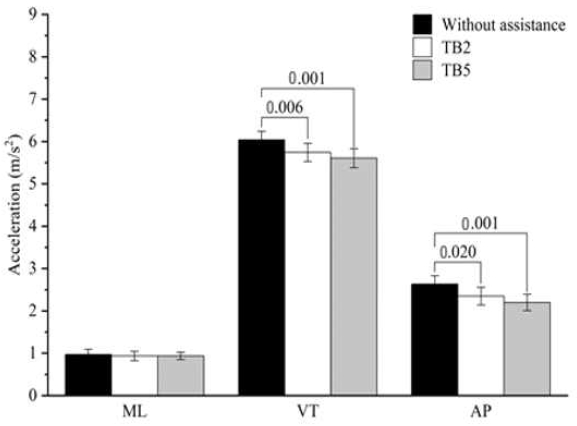
Significant differences were observed in VT (p = 0.006) and AP (p = 0.020) movements between walking without assistance and with TB2. Furthermore, significant differences were observed in VT (p = 0.001) and AP (p = 0.001) movements between walking without assistance and with TB5. No significant differences were observed in AP and VT movement between walking with TB2 and walking with TB5 (p > 0.05). The ML-direction acceleration values did not differ significantly among walking without assistance, with TB2, and with TB5 (p > 0.05).
The application of the newly developed weighted theraband banding can improve anterior-posterior and vertical movements of the thoracic spine. It is believed to have pulled the thoracic spine to enhance stability during walking, thereby reducing anterior-posterior and vertical movement in subjects with thoracic kyphosis.
Keywords:
3-axis accelerometer, Theraband, Thoracic Kyphosis, Thoracic MovementⅠ. Introduction
Since the 1950s, computer and mobile technologies have revolutionized human society and employment (Egger et al, 2001). In the workplace, employees often adopt a slouched sitting posture during breaks (Watanabe et al, 2007), which may lead to lumbar extensor muscle fatigue and difficulty maintaining the spine in a normal upright posture (Carter and Banister, 1994). Prolonged slouched posture can cause spinal misalignment, resulting in thoracic kyphosis (TK), rounded shoulder posture, or forward head posture (Chansirinukor et al, 2001; Magee, 2002). The use of inappropriate tables and chairs also contributes to abnormal posture among individuals who sit for extended periods, such as office workers and primary and middle school students. Additionally, the use of heavy backpacks or engagement in activities that cause back pressure can lead to spinal misalignment (Britnell et al, 2005).
Sagittal spinal alignment is primarily influenced by the thoracic spine, and the thoracic curve is determined by the shape of the vertebral bodies (Macagno and O’Brien, 2006). The thoracic vertebrae have limited mobility, thereby preventing damage to the nerve trunks and abdominal viscera (Lee, 2010). A normal thoracic angle is typically < 40° (Neumann, 2010), whereas an angle > 40° indicates the presence of TK (Eum et al, 2013). Prolonged TK can result in narrowing of the intervertebral spaces; compression of the heart (Katzman et al, 2015); restricted air entry into the thoracic cavity leading to an abnormal, shallow, and interrupted breathing pattern (Leech et al, 1990); increased risk of spinal fractures (McGrother et al, 2002); back pain; disability (Ettinger et al, 1994); and decreased quality of life (Martin et al, 2002). Several studies have focused on musculoskeletal management of TK. For example, some studies have investigated the effects of pectoralis major muscle stretching exercises on TK to achieve muscle stretching and relaxation, as well as the impact of scapular stabilization training on sagittal spinal alignment. In 2016, Im et al. (2016) conducted a study in which participants were randomly assigned to a scapular stabilization group or a control group. In the scapular stabilization group, intensive scapular stabilization training was associated with reduced neck pain and improved sagittal spinal alignment. Additionally, Jeong and Kim (2019) evaluated the effect of thoracic joint mobilization on changes in the TK angle. Numerous studies have explored TK and its treatment methods, including chest stretching exercises, scapular stabilization, draft training, and intensive training of various related muscles (Eum et al, 2013; Jeong and Kim, 2019; Im et al, 2016; Katzman et al, 2015).
Theraband is a pioneer technology developed for progressive resistance training, with proven effectiveness in improving muscle strength, mobility, motor function, and joint pain (Macagno and O’Brien, 2006). Additionally, it can facilitate isometric contractions of muscles prior to activation or mutual inhibition during therapeutic exercises (Greendale et al, 2011). A previous study categorized young individuals with TK into self-activity and intensive exercise groups (Lee et al, 2019). The intensive exercise regimen involved the use of theraband and dumbbells for resistance training of the extensor dorsalis muscle. Compared with the self-activity group, the intensive exercise group demonstrated significant improvements in TK. However, no studies have evaluated movement of thoracic region after applying theraband banding in the TK group. It is important to evaluate the movement of the thoracic region before and after intervention, as large movements of the thoracic in patients with excessive TK can increase instability of the trunk.
In the present study, we optimized the correction potential of theraband through a novel approach inspired by weighted orthotics. This approach involved affixing sandbags onto theraband; the resulting weighted theraband banding. We hypothesized that weighted theraband banding would have some effect on the thoracic movement in individuals with TK. Therefore, this study aimed to develop the weighted theraband banding and explore its effects on TK movement.
Ⅱ. Methods
1. Subjects
This study included 14 volunteers (7 men and 7 women) aged 30-60 years with TK angle ≥ 40° who had no contraindications to exercise, such as rheumatic disease, spinal arthritis, or nervous system disease (Song et al, 2013), history of spinal surgery (Jeong and Kim, 2019), contact dermatitis, or skin reaction to kinematic tapes (Zhu et al, 2017) or theraband treatment (Yun et al, 2020). The study protocol was approved by the institutional review board, and all participants provided written informed consent. The participants had a mean age of 42.3 ± 5.2 years, mean height of 171.4 ± 3.4 cm, mean weight of 64.5 ± 7.1 kg, and mean TK angle of 43.4 ± 3.0°. We received an informed consent from all participants for all information about the experiment.
2. Equipments
TK angles were measured using a Dual Inclinometer (Acumar, Lafayette Instrument Co., Lafayette, IN, USA) (Shin et al, 2016). First, the T1 and T12 spinous processes of the participants were marked. Then, with the volunteers standing comfortably and looking straight ahead, the two ends of the dual inclinometer were placed at the two marks; the incline was recorded as the TK angle. This process was repeated three times, and the mean value was recorded to minimize error.
A 3-axis accelerometer (Fit dot life, Suwon, Korea; size: 35 × 35 × 13 mm; weight: 13.7 g) was fixed at the level of the spinous process of the sixth thoracic vertebra to measure the changes in TK in the anterior-posterior (AP), vertical (VT), and medial-lateral (ML) directions in the frontal plane at 30 Hz (Patil and Rao, 2011). The data were calculated with the following equations and the value indicate the movement in the AP, VT, and ML directions (Liu and Yoo, 2022). Measurements were collected at a sampling rate of 30 Hz, that is, 30 acceleration values are recorded every second. The data were calculated with the following equations, “n” represents the number of acceleration values in the corresponding movement time, but there are positive and negative acceleration values in the movement. We calculate the positive number of each acceleration value by , and then calculate the average acceleration value in the corresponding time period. The calculated values indicate the movement (oscillation) of each direction.
Theraband (Thera-Band®, Hygenic Corp, Akron, USA) is available in tea, yellow, red, green, blue, black, silver, and gold colors, which indicate increasing elastic resistance. Considering that red is the international standard color of theraband, which provides 3.7 pounds of resistance at 100% elongation (Shin et al, 2018), the red theraband was used for training in this study. During cutting and fixing of the theraband, participants were required to sit comfortably with their upper limbs stretched out on their backs and their hands clasped to contract both shoulder blades. Then, the head was retracted to the neutral position, and the distance between the acromion on both sides was measured using a tape measure. This distance represents the true distance between the acromion joints in each participant. The length of the elastic belt was set to half the distance between the acromion joints, yielding 100% elastic resistance. Appropriate knotted lengths were reserved at both ends. Using the aforementioned method, two elastic belts were cut.
We set the applied weight to 2% and 5% of each participant’s body weight using a sandbag. The sandbags had drawstrings that could be used to alter sand capacity and weight according to the participant’s body weight. Based on the mean participant height, the sandbags were designed with a length and width of 17 and 15 cm, respectively.
3. Experimental methods
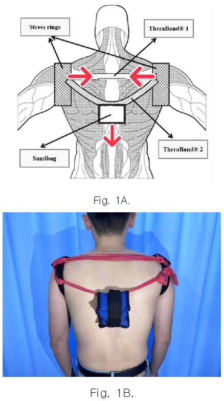
A 3-m straight line was marked on a flat ground. To achieve the natural walking state, participants were instructed to walk from 1 m before to 1 m after the marked 3-m line (i.e., a total of 5 m). However, data were collected only while participants walked on the 3-m line. First, the three-axis accelerometer was fixed at the level of the spinous process of the sixth thoracic vertebra using double-sided adhesive tape. Next, data were collected during the unassisted 3-m walk, and the mean value from three repetitions was recorded. Then, stress rings were securely fixed at both of the participant’s shoulder joints, and two therabands were fixed between the two stress rings. A sandbag weighing 2% of the participant’s body weight was attached to the midpoint of a theraband (TB2)(Fig. 1). Participants were instructed to walk for 3 m three times, and the mean value was recorded. To prevent muscle fatigue and tension, participants were allowed to rest for 30 s between trials. Subsequently, the sandbags were replaced with sandbags weighing 5% of the participant’s body weight (TB5), and the process was repeated.
4. Data analysis
Data were analyzed using SPSS software (version 20.0; IBM Corp., Armonk, NY, USA). The effect size calculated by G-Power was 0.89, exceeding the minimum efficacy threshold of 0.8. The variables displayed a normal distribution. Therefore, one-way repeated measures analysis of variance was used to identify significant differences in the change in acceleration of the thoracic vertebra in the AP, VT, and ML directions.
Ⅲ. Results
1. AP movements
Table 1 presents the acceleration values in the AP direction. The AP movements were significantly different between walking without assistance and walking with TB2 (p < 0.05) and between walking without assistance and walking with TB5 (p < 0.05). No significant differences were observed in AP movement between walking with TB2 and walking with TB5 (p > 0.05)(Fig.2).

Acceleration values in the anterior-posterior (AP), vertical (VT), and medial-lateral (ML) directions(N = 14)
2. VT movements
The acceleration values are shown in Table1. There were significant differences between walking without assistance and walking with TB2 (p < 0.05) and between walking without assistance and walking with TB5 (p < 0.05). There were no statistically significant differences in VT movement between walking with TB2 and walking with TB5 (p > 0.05)(Fig.2).
3. ML movements
Table 1 presents the acceleration values in the ML direction. These acceleration values were not significantly different among walking without assistance, with TB2, and with TB5 (p > 0.05). No significant differences were observed in ML movements of the thoracic vertebrae before and after using the weighted theraband banding (Fig. 2).
Ⅳ. Discussion
Compared to walking without assistance, walking with the assistance of TB2 or TB5 was associated with significantly reduced VT and AP thoracic movements. Although no significant differences were observed between walking with the assistance of TB2 and walking with the assistance of TB5 (p > 0.05), TB5 was associated with better control of thoracic vertebral movements, as demonstrated by smaller acceleration values in all directions.
Participants with TK had larger movements in the AP and VT directions when walking without assistance compared to walking with weighted theraband banding assistance, which may be explained by the greater trunk flexion during walking without assistance. Previous studies have demonstrated forward deviation of the center of gravity among individuals with TK compared to individuals without TK, leading to increased thoracic bending torque (Neumann, 2010), imbalance, and fall risk (Eum et al, 2013).
During walking, the normal spinal curvature compensates for the impact from the ground reaction, leading to a VT movement of almost 5 cm (Patil and Rao, 2011). TK is characterized by an excessively inverted “C”-shape of the thoracic spine, which may be associated with lumbar kyphosis and reduced spinal capacity to cushion the impact. In individuals with TK, the ground reaction force is transmitted directly to the spine during walking, particularly on stairs, thereby aggravating trunk instability and leading to larger VT movements of the center of mass (Mirafzal et al, 2011). In the present study, compared to walking with theraband banding assistance, walking without assistance was associated with larger thoracic spine movements in the AP and VT directions, which partially compensated for the thoracic spine instability. To overcome spinal instability, the human body adopts an optimal position for maintaining the center of gravity. This process is associated with substantial increases in AP and VT acceleration of the thoracic spine during walking and climbing stairs, which ensure rapid adjustment of the center of gravity.
We fixed two therabands at 100% elongation between both shoulders, simulating the application of passive traction to the abnormal scapula that rotates inward and abducts, to correct the passive outward rotation and adduction of the scapula. Additionally, sandbags of different weights were fixed to a theraband to simulate the application of external passive backward traction to the abnormally shaped thoracic spine. We found that the weighted theraband banding effectively reduced the AP and VT movements, suggesting that the application of passive traction forces by the weighted theraband banding can reduce excessive thoracic curvature and unnecessary movement of the thoracic spine.
In this study, we used the elasticity and mechanical load of the weighted theraband to facilitate effective correction of TK. Participants walked for 3 m using a theraband with 100% elongation fixed by weighted sandbags. During walking, the sandbag fixed at the midpoint of the theraband was elastically pulled by gravity, maximizing the backward pull among individuals with TK. Additionally, the weight of the sandbags was determined according to the body weight, facilitating personalized treatment of TK. The AP and VT movements were significantly reduced during dynamic activity when applying the weighted theraband banding. The newly developed weighted theraband banding technique is expected to enhance load assistance, allowing its application during dynamic activity. The present study had several limitations. First, it only included 14 participants, limiting the generalizability of the findings. Nevertheless, the participants had a wide age range. Second, we did not evaluate the long-term effects of weighted theraband banding on TK. Further studies with larger sample sizes are needed to evaluate the long-term effect and to compare effectiveness between the original theraband technique and weighted theraband banding.
Ⅴ. Conclusion
The application of the newly developed weighted theraband banding can improve the anterior-posterior and vertical movements of the thoracic vertebra by rapidly correcting the abnormal positions of the scapula and cervicothoracic line. Furthermore, it is believed to have pulled the thoracic spine to provide stability during walking, thereby reducing the movements of anterior-posterior and vertical in subjects with thoracic kyphosis.
References
-
Britnell SJ, Cole JV, Isherwood L, et al. Postural health in women: the role of physiotherapy. J Obstet Gynaec Can 2005;27:493-510.
[https://doi.org/10.1016/S1701-2163(16)30535-7]

-
Carter JB, Banister EW. Musculoskeletal problems in VDT work: a review. Ergonomics 1994;37:1623-1648.
[https://doi.org/10.1080/00140139408964941]

-
Chansirinukor W, Wilson D, Grimmer K, et al. Effects of backpacks on students: measurement of cervical and shoulder posture. Aust J Physiother 2001;47:110-116.
[https://doi.org/10.1016/S0004-9514(14)60302-0]

-
Egger M, Davey Smith G, Altman DG. Systematic reviews in health care. London: BMJBooks; 2001.
[https://doi.org/10.1002/9780470693926]

-
Ettinger B, Black DM, Palermo L, et al. Kyphosis in older women and its relation to back pain, disability and osteopenia: the study of osteoporotic fractures. Osteoporos Int 1994;4:55-60.
[https://doi.org/10.1007/BF02352262]

-
Eum R, Leveille SG, Kiely DK, et al. Is kyphosis related to mobility, balance and disability? Am J Phys Med Rehabil 2013;92:980-989.
[https://doi.org/10.1097/PHM.0b013e31829233ee]

-
Greendale GA, Nili NS, Huang MH, et al. The reliability and validity of three non-radiological measures of thoracic kyphosis and their relations to the standing radiological Cobb angle. Osteoporos Int 2011;22:1897-1905.
[https://doi.org/10.1007/s00198-010-1422-z]

-
Im B, Kim Y, Chung Y, et al. Effects of scapular stabilization exercise on neck posture and muscle activation in individuals with neck pain and forward head posture. J Phys Ther Sci 2016;28:951-955.
[https://doi.org/10.1589/jpts.28.951]

-
Jeong HJ, Kim BJ. The effect of thoracic joint mobilization on the changes of the thoracic kyphosis angle and static and dynamic balance. Biomed Sci Lett 2019;25:149-158.
[https://doi.org/10.15616/BSL.2019.25.2.149]

-
Katzman WB, Harrison SL, Fink HA, et al. Physical function in older men with hyperkyphosis. J Gerontol A Biol Sci Med Sci 2015;70:635-640.
[https://doi.org/10.1093/gerona/glu213]

- Lee HA. A study on the safety of spinal manipulation using finite element method [a master's thesis]. Korea University; 2010.
-
Lee JH, Jeon HS, Kim JH, et al. Immediate effects of the downhill treadmill walking exercise on thoracic angle and thoracic extensor muscle activity in subjects with thoracic kyphosis. Phys Ther Korea 2019;26:1-7.
[https://doi.org/10.12674/ptk.2019.26.2.001]

-
Leech JA, Dulberg C, Kellie S, et al. Relationship of lung function to severity of osteoporosis in women. Am Rev Respir Dis 1990;141:68-71.
[https://doi.org/10.1164/ajrccm/141.1.68]

-
Liu YY, Yoo WG. Effects of lower trunk movement in flat-back syndrome during stair climbing: a technical note. Technol Health Care 2022;30:483-489.
[https://doi.org/10.3233/THC-202668]

-
Macagno AE, O’Brien MF. Thoracic and thoracolumbar kyphosis in adults. Spine (Phila Pa 1976) 2006;31:161-170.
[https://doi.org/10.1097/01.brs.0000236909.26123.f8]

- Magee DJ. Orthopedic Physical Assessment. 2nd ed. Philadelphia: Saunders; 2002.
-
Martin AR, Sornay-Rendu E, Chandler JM, et al. The impact of osteoporosis on quality-of-life: the OFELY cohort. Bone 2002;31:32-36.
[https://doi.org/10.1016/S8756-3282(02)00787-1]

-
McGrother CW, Donaldson MM, Clayton D, et al. Evaluation of a hip fracture risk score for assessing elderly women: the Melton Osteoporotic Fracture (MOF) study. Osteoporos Int 2002;13:89-96.
[https://doi.org/10.1007/s198-002-8343-6]

- Mirafzal S, Sokhanguee Y, Sadeghi H. The effect of a combination of corrective exercise and spinal taping on balance in kyphotic adolescent. SSQ 2011;2:18-24.
- Neumann DA. Kinesiology of the musculoskeletal system: foundations for physical rehabilitation. St Louis: Mosby; 2010.
- Patil P, Rao S. Effects of Thera-Band® elastic resistance-assisted gait training in stroke patients: a pilot study. Eur J Phys Rehabil Med 2011;47:427-433.
-
Shin AR, Lee JH, Kim DE, et al. Thera-Band application changes muscle activity and kyphosis and scapular winging during knee push-up plus in subjects with scapular winging. The cross-sectional study. Medicine (Baltimore) 2018;97:e0348.
[https://doi.org/10.1097/MD.0000000000010348]

-
Shin S, An D. Yoo W. Effects of differences in visual acuity on gait time and trunk acceleration when older women negotiate stairs. Eur Geriatr Med 2016;7:538-542.
[https://doi.org/10.1016/j.eurger.2016.04.003]

- Song JE, Kim SY, Jang HJ. A comparison of the effects of self-mobilization and strengthening exercise of the thoracic region in young adults with thoracic hyperkyphosis. J Korean Orthop Manu Phys Ther 2013;19:11-18.
- Watanabe S, Eguchi A, Kobara K, et al. Influence of trunk muscle co-contraction on spinal curvature during sitting for desk work. Electromyogr Clin Neurophysiol 2007;47:273-278.
-
Yun HG, Lee JH, Choi IR. Effects of kinesiology taping on shoulder posture and peak torque in junior baseball players with rounded shoulder posture: a pilot study. Life (Basel) 2020;10:139.
[https://doi.org/10.3390/life10080139]

-
Zhu C, Yang X, Zhou B, et al. Cervical kyphosis in patients with Lenke type 1 adolescent idiopathic scoliosis: the prediction of thoracic inlet angle. BMC Musculoskelet Disord 2017;18:220.
[https://doi.org/10.1186/s12891-017-1590-5]


