
Electromyographic Analysis of Shoulder External Rotators and Scapular Stabilizers During Eccentric Shoulder External Rotation at Different Shoulder Abduction Angles
© 2026 by the Korean Physical Therapy Science
Abstract
Shoulder external rotators and scapular stabilizers play important roles in maintaining glenohumeral stability during overhead activities. Although eccentric shoulder external rotation (ESER) is clinically important in overhead athletes, muscle activation patterns during ESER remain unclear at different shoulder abduction angles.
Cross-sectional study.
Thirty healthy male participants voluntarily participated in this study. ESER strength and surface electromyography (sEMG) data were collected under four shoulder abduction conditions (0°, 45°, 90°, and 135° in the scapular plane). Muscle activity of the infraspinatus, supraspinatus, serratus anterior, upper trapezius, middle trapezius, and lower trapezius was measured and normalized to maximal voluntary isometric contraction (%MVIC). One-way repeated-measures analysis of variance with Bonferroni correction was used to compare variables among shoulder abduction angles.
No significant differences in ESER strength were observed among shoulder abduction angles (p>0.05). Infraspinatus activity significantly decreased as shoulder abduction angle increased (p<0.05). In contrast, supraspinatus and serratus anterior activity significantly increased at higher shoulder abduction angles (p<0.05). Upper trapezius activity also increased significantly with increasing shoulder abduction angle (p<0.05).
Shoulder abduction angle altered muscle recruitment during ESER without changing external rotation strength. Lower angles may emphasize infraspinatus activation, whereas higher angles may increase supraspinatus and serratus anterior recruitment. These findings may help clinicians select ESER positions according to the target muscle and rehabilitation goal.
Keywords:
Electromyography, Muscle contraction, Muscle strength, Rotator cuff, ShoulderⅠ. Introduction
The shoulder requires coordinated glenohumeral and scapulothoracic motion to balance mobility and stability. The rotator cuff muscles contribute to humeral head stabilization, while the trapezius and serratus anterior support scapular positioning and dynamic shoulder stability (Halder et al., 2001; Sahrmann, 2002). Altered activation of scapular stabilizers has been associated with abnormal scapular kinematics and shoulder dysfunction (Paine & Voight, 2013).
Shoulder disorders are common in both athletes and the general population, and repetitive overhead activity is considered a major risk factor for shoulder injury (Luime et al., 2004; McFarland & Wasik, 1998). In overhead sports, the deceleration phase of throwing places substantial eccentric demand on the shoulder external rotators, which function as dynamic decelerators (Cools et al., 2015). Repetitive eccentric loading may contribute to overuse injuries and muscular imbalance between the shoulder internal and external rotators (Fleisig et al., 1995). Previous studies have suggested that insufficient eccentric strength of the external rotators is associated with an increased risk of shoulder injury in overhead athletes (Niederbracht et al., 2008).
Eccentric strengthening exercises have been reported to improve muscular imbalance of the shoulder rotators more effectively than concentric exercises (Niederbracht et al., 2008). Consequently, eccentric training and assessment of the shoulder external rotators are commonly incorporated into rehabilitation and return-to-sport programs for overhead athletes (Kovacic & Bergfeld, 2005). Although several studies have investigated muscle activation during concentric or isometric shoulder external rotation at different shoulder abduction angles (Reinold et al., 2004; Uga et al., 2016), little is known about muscle activation patterns during eccentric shoulder external rotation (ESER), which more closely reflects the functional demands of overhead sports.
Recent evidence indicates that shoulder muscle recruitment during external rotation is influenced by forearm position, body position, humeral abduction angle, and exercise resistance. Hedt et al. (2020) demonstrated that forearm position altered muscle recruitment during several eccentric shoulder exercises. Whittaker et al. (2022) reported region-specific activation of the infraspinatus and supraspinatus across different arm postures during isometric external rotation. Sung et al. (2023) further showed that upper trapezius and serratus anterior activity was greater at 90° than at 45° of shoulder abduction during external rotation exercise. More recently, Sung et al. (2026) reported angle-dependent changes in supraspinatus, upper trapezius, and serratus anterior activity during elastic-band external rotation exercises.
However, previous studies have primarily examined isometric or elastic-resistance tasks, compared a limited number of abduction angles, or evaluated different eccentric exercise variations rather than the same standardized eccentric task across a wide range of shoulder positions. Therefore, it remains unclear whether ESER strength and the coordinated activity of the shoulder external rotators and scapular stabilizers change systematically from 0° to 135° of shoulder abduction. The present study simultaneously compared ESER strength and the activity of the infraspinatus, supraspinatus, serratus anterior, and three regions of the trapezius at 0°, 45°, 90°, and 135° in the scapular plane. By maintaining the same contraction mode and movement direction while varying the abduction angle, this study aimed to isolate angle-dependent muscle recruitment during eccentric loading.
Ⅱ. Methods
1. Study Design
This study employed a cross-sectional study design to investigate muscle activation patterns during eccentric shoulder external rotation (ESER) at different shoulder abduction angles.
2. Participants
Thirty healthy adult males voluntarily participated in this study. Participants were recruited from S University located in Seoul, Republic of Korea. Inclusion criteria were healthy males younger than 30 years of age. Exclusion criteria included a history of neck or shoulder pain within the previous 6 months, previous surgery involving the neck or shoulder, current symptoms related to the neck or shoulder, passive shoulder abduction range of motion less than 135°, or passive shoulder external rotation range of motion less than 90° (Papotto et al., 2016).
Sample size estimation was performed using G*Power software based on the infraspinatus muscle activity reported in a previous study by Uga et al. (2016). The minimum required sample size was calculated as seven participants. Considering the generalizability and statistical stability of the results, a total of 30 participants were recruited.
All participants received a detailed explanation regarding the purpose and procedures of the study before participation and provided written informed consent. This study was approved by the Institutional Review Board of Sahmyook University (approval No. 2-7001793-AB-N-012019001HR). The general characteristics of the participants are presented in Table 1.

General characteristics of the participants(N=30)
3. Experimental Procedures
Participants performed ESER under four shoulder abduction conditions (0°, 45°, 90°, and 135°) in the scapular plane, defined as 30° anterior to the frontal plane (Kramer & Ng, 1996). Participants were seated on a chair with the trunk stabilized using a belt to minimize compensatory trunk movement. The elbow joint was maintained at 90° of flexion throughout the experiment. At 0° of shoulder abduction, the examiner supported the participant’s elbow. At 45°, 90°, and 135° of shoulder abduction, the distal humerus was supported on an adjustable treatment table to minimize the muscular effort required to maintain the shoulder abduction position (Uga et al., 2016). The experimental positions for each shoulder abduction condition are shown in Figure 1.
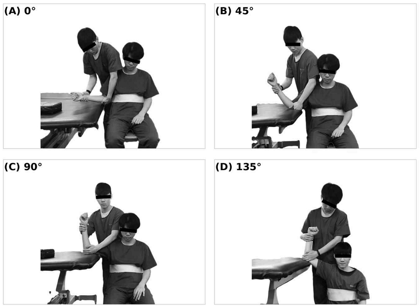
Experimental positions for eccentric shoulder external rotation at shoulder abduction angles of (A) 0°, (B) 45°, (C) 90°, and (D) 135° in the scapular plane. Participants were seated with the trunk stabilized using a belt.
To induce eccentric contraction of the shoulder external rotators, participants were instructed to exert maximal shoulder external rotation effort while the examiner manually moved the arm from maximal external rotation toward the neutral position, following the protocol described by Johansson et al. (2015). A metronome was used to maintain a consistent movement speed during testing.
Each condition was performed for 5 seconds and repeated three times. A 30-second rest period was provided between trials to minimize fatigue. Testing was discontinued if compensatory movements were observed and resumed after an appropriate rest period.
4. Outcome Measures
Shoulder external rotation strength was measured using a hand-held dynamometer (HHD; Lafayette Instrument, Lafayette, IN, USA). The HHD was positioned on the dorsal aspect of the forearm, approximately 2 cm proximal to the ulnar styloid process. Peak force values measured in kilograms were converted to Newtons (N) by multiplying by gravitational acceleration (9.81 m/s²). Three trials were performed for each condition, and the average value was used for analysis.
The standardized eccentric HHD procedure used in the present study was based on the method described by Johansson et al. (2015). In their study, this procedure demonstrated excellent intratester reliability (ICC=0.858–0.879), good intertester reliability (ICC=0.714), and good concurrent validity with isokinetic dynamometry (r=0.70–0.78).
Surface electromyography (sEMG) data were collected using the TeleMyo 2400T G2 Telemetry EMG system (Noraxon Inc., Scottsdale, AZ, USA). Muscle activity was recorded from the infraspinatus, supraspinatus, serratus anterior, upper trapezius, middle trapezius, and lower trapezius muscles.
Before electrode placement, the skin was shaved and cleaned with alcohol to reduce skin impedance. Electrode placement followed the methods described by Criswell and Cram (2011). Electromyographic signals were sampled at 1,000 Hz with a band-pass filter of 20–450 Hz and a 60 Hz notch filter. Raw EMG signals were processed using the root mean square method.
To normalize muscle activity, maximal voluntary isometric contraction (MVIC) values were collected according to the manual muscle testing procedures described by Kendall et al. (2005). All EMG data were normalized to MVIC and expressed as percentage MVIC (%MVIC). The middle 3 seconds of each 5-second trial were used for data analysis.
5. Statistical Analysis
All statistical analyses were performed using IBM SPSS Statistics version 22.0 (IBM Corp., Armonk, NY, USA). Descriptive statistics were calculated for participant characteristics. One-way repeated-measures analysis of variance (ANOVA) was used to compare differences in muscle activity and strength among shoulder abduction angles. Bonferroni correction was applied for post-hoc analysis. Statistical significance was set at p<0.05.
Ⅲ. Results
1. Shoulder External Rotation Strength
The ESER strength measured using a hand-held dynamometer under different shoulder abduction angles is presented in Table 2. Mean ESER strength values were 128 N, 133 N, 134 N, and 130 N at shoulder abduction angles of 0°, 45°, 90°, and 135°, respectively. No significant differences in ESER strength were observed among shoulder abduction angles (p>0.05).
Eccentric shoulder external rotation strength according to shoulder abduction angle(N=30)
2. Muscle Activity
Normalized muscle activity during ESER at different shoulder abduction angles is presented in Table 3.
Muscle activation during eccentric shoulder external rotation according to shoulder abduction angle(N=30)
Infraspinatus activity significantly decreased as shoulder abduction angle increased (p<0.05). In contrast, supraspinatus activity significantly increased with increasing shoulder abduction angle (p<0.05).
Serratus anterior and upper trapezius activities significantly increased as shoulder abduction angle increased (p<0.05). In contrast, middle trapezius activity significantly decreased at higher shoulder abduction angles (p<0.05).
Lower trapezius activity also showed a decreasing trend as shoulder abduction angle increased, with significantly lower activation observed at 90° and 135° compared with lower shoulder abduction angles (p<0.05).
Ⅳ. Discussion
The purpose of this study was to investigate the activation patterns of the shoulder external rotators and scapular stabilizing muscles during eccentric shoulder external rotation (ESER) at different shoulder abduction angles. The results demonstrated that ESER strength did not differ significantly among the four shoulder abduction conditions, whereas muscle activation patterns changed considerably according to shoulder position. Specifically, infraspinatus activity decreased as the shoulder abduction angle increased, whereas supraspinatus, serratus anterior, and upper trapezius activity increased. In contrast, middle and lower trapezius activity decreased at higher shoulder abduction angles. These findings indicate that similar external rotation force output may be produced through different combinations of rotator cuff and scapular muscle recruitment depending on shoulder position.
The infraspinatus is considered a primary external rotator of the glenohumeral joint across different shoulder positions (Kuechle et al., 2000). In the present study, infraspinatus activity progressively decreased as the shoulder abduction angle increased. This pattern is generally consistent with previous studies investigating concentric and isometric shoulder external rotation exercises (Reinold et al., 2004; Uga et al., 2016). Reinold et al. (2004) reported that infraspinatus activity differed among external rotation exercises performed in different arm positions, while Uga et al. (2016) observed lower infraspinatus activity at higher shoulder elevation angles during isometric external rotation. These findings suggest that lower shoulder abduction positions may place the infraspinatus in a more favorable position for producing axial external rotation, whereas elevation of the humerus may alter its muscle length, line of action, and mechanical contribution to the task.
However, the infraspinatus is not functionally homogeneous, and activation may differ among its anatomical regions. Whittaker et al. (2022), using fine-wire electromyography, reported that the superior, middle, and inferior regions of the infraspinatus did not respond uniformly to changes in arm posture. In particular, inferior infraspinatus activity increased in some elevated arm positions, whereas the activities of the superior and middle regions were less sensitive to posture. This finding does not entirely correspond with the progressive reduction in whole-muscle surface EMG observed in the present study. The apparent difference may be attributable to differences in electrode type, contraction mode, exercise plane, and regional sampling. Because surface EMG primarily represents the activity of the superficial muscle fibers beneath the electrodes (Criswell & Cram, 2011), the decreased infraspinatus activity observed in the present study may reflect a reduction in the recorded superficial region rather than a uniform reduction across the entire muscle. Therefore, the present results are more appropriately interpreted as evidence of angle-dependent redistribution of rotator cuff recruitment rather than a reduction in the overall functional contribution of the infraspinatus.
In contrast to the infraspinatus, supraspinatus activity increased significantly as the shoulder abduction angle increased. Although the supraspinatus is commonly recognized for its contribution to shoulder abduction, it also contributes to glenohumeral compression, stabilization, and external rotation (Veeger et al., 1991; Tardo et al., 2013). As the humerus is elevated, the glenohumeral joint may require greater compressive and stabilizing forces to maintain the humeral head within the glenoid while external rotation force is produced. Therefore, the increased supraspinatus activity at higher abduction angles may reflect greater demand for glenohumeral stabilization as well as an increased contribution to external rotation. Whittaker et al. (2022) similarly reported that the anterior and posterior regions of the supraspinatus were more active during external rotation performed at 90° of abduction or scaption than during several lower-elevation positions. More recently, Sung et al. (2026) found that supraspinatus activity increased at 45° and 90° of shoulder abduction compared with 0° during elastic-resistance external rotation. Taken together, these findings suggest that elevation of the humerus changes the relative contributions of the infraspinatus and supraspinatus, increasing the stabilizing and external rotation demands placed on the supraspinatus at higher abduction angles.
Serratus anterior and upper trapezius activity also increased as the shoulder abduction angle increased. These muscles function cooperatively to control scapular upward rotation and positioning during arm elevation. The serratus anterior additionally contributes to posterior tilting and external rotation of the scapula, thereby helping maintain favorable glenoid alignment and subacromial space during elevated arm positions (Paine & Voight, 2013; McClure et al., 2006). Therefore, the increased serratus anterior activity observed in the present study may reflect greater demand for scapular stabilization and posterior tilting as the humerus was positioned at higher abduction angles. Increased upper trapezius activity may similarly represent a greater requirement for scapular upward rotation and elevation. Recent studies have demonstrated that shoulder position influences serratus anterior and upper trapezius recruitment during shoulder exercises (Sung et al., 2023; Jang et al., 2026; Sung et al., 2026). These findings support the interpretation that higher shoulder abduction angles increase the contribution of the upper trapezius–serratus anterior force couple during external rotation exercises.
The reduction in middle and lower trapezius activity at higher shoulder abduction angles should be interpreted more cautiously. One possible explanation is that the scapular control strategy shifted toward greater reliance on the upper trapezius and serratus anterior as the humerus was elevated, with relatively less recruitment of the middle and lower trapezius during the specific eccentric external rotation task used in this study. The middle trapezius primarily contributes to scapular retraction and stabilization, whereas the lower trapezius assists scapular stabilization, posterior tilting, and upward rotation (Paine & Voight, 2013); moreover, the relative recruitment of the upper and lower trapezius may vary according to exercise configuration (Kim, 2026). In addition, because the distal humerus was supported at the elevated shoulder positions, the support surface may have reduced some of the muscular demand associated with maintaining the arm position. Changes in muscle length and the relationship between the muscle fibers and surface electrodes at different scapular positions may also have influenced the recorded EMG amplitude. Because scapular kinematics and muscle lengths were not measured directly, these explanations remain plausible mechanisms rather than confirmed causal relationships.
Another notable finding was that measured ESER strength remained relatively constant despite substantial changes in muscle activity. This result suggests that the shoulder complex may use different recruitment combinations to maintain similar external rotation force output across abduction angles. The decreased surface EMG activity of the infraspinatus at higher abduction angles may have been accompanied by greater activation of the supraspinatus and scapular stabilizers. However, the present data do not demonstrate direct compensation by one muscle for another. Muscles that were not measured, including the teres minor and posterior deltoid, may also have contributed to the measured external rotation force. Thus, the results are best interpreted as showing angle-dependent redistribution of muscle recruitment while the overall external rotation strength remained similar.
From a clinical perspective, these findings suggest that shoulder abduction angle may be adjusted according to the muscle targeted and the stage of rehabilitation. External rotation performed at lower shoulder abduction angles may be useful when greater infraspinatus activation is desired with comparatively lower supraspinatus, serratus anterior, and upper trapezius involvement. In contrast, external rotation at higher abduction angles may provide a greater challenge to the supraspinatus and the upper trapezius–serratus anterior force couple and may therefore be more appropriate during later rehabilitation stages or sport-specific preparation for overhead activity. Nevertheless, greater EMG activity should not automatically be interpreted as a superior therapeutic effect. Exercise position should be selected according to tissue healing, symptoms, movement quality, and the intended rehabilitation goal. Furthermore, because the participants were healthy young men rather than overhead athletes or patients with shoulder disorders, the clinical implications should be applied cautiously.
This study has several limitations. First, the participants were limited to healthy young adult males, which restricts the generalizability of the findings to female participants, older adults, overhead athletes, and individuals with shoulder pathology. Second, surface EMG was used to assess muscle activity. Consequently, deep muscles such as the teres minor could not be evaluated, and the recordings could not distinguish activation among the anatomical regions of the infraspinatus and supraspinatus. Surface EMG measurements may also have been influenced by cross-talk and by changes in muscle length or electrode orientation across shoulder positions. Third, eccentric movement was imposed manually by the examiner while the participants resisted maximally. Although a metronome, standardized movement duration, and consistent verbal instructions were used, the magnitude and direction of the manual resistance and the examiner’s ability to maintain a constant movement velocity may have varied among participants, trials, and shoulder positions. Examiner leverage and stabilization of the hand-held dynamometer may also have differed according to the abduction angle, potentially affecting both the strength and EMG outcomes. Fourth, scapular kinematics and glenohumeral joint motion were not measured directly. Therefore, the proposed explanations involving scapular upward rotation, posterior tilting, muscle length, and joint stabilization represent biomechanically plausible interpretations rather than mechanisms directly demonstrated by the present study. Future studies should use externally fixed dynamometry or an isokinetic dynamometer to standardize resistance and angular velocity, incorporate three-dimensional scapular kinematic measurements, and examine regional rotator cuff activity using fine-wire EMG. Studies involving overhead athletes and individuals with shoulder disorders are also required to determine whether the observed recruitment patterns are clinically reproducible.
Ⅴ. Conclusion
Muscle activation patterns during eccentric shoulder external rotation varied according to shoulder abduction angle despite similar external rotation strength among conditions. Infraspinatus activity decreased as shoulder abduction angle increased, whereas supraspinatus and serratus anterior activity increased at higher shoulder abduction angles. These findings suggest that different muscular recruitment strategies may contribute to maintaining similar external rotation force output across shoulder positions. Clinically, shoulder abduction angle may be considered when selecting eccentric shoulder external rotation exercises according to the target muscle and rehabilitation goal, although confirmation in overhead athletes and patients with shoulder disorders is needed.
Acknowledgments
This paper was supported by the Sahmyook University Research Fund in 2025 (RI12025031).
References
-
Cools AM, Johansson FR, Borms D, et al. Prevention of shoulder injuries in overhead athletes: a science-based approach. Braz J Phys Ther 2015;19(5):331-339.
[https://doi.org/10.1590/bjpt-rbf.2014.0109]

- Criswell E, Cram JR. Cram’s introduction to surface electromyography. 2nd ed. Burlington: Jones and Bartlett Publishers; 2011. p.289-302.
-
Fleisig GS, Andrews JR, Dillman CJ, et al. Kinetics of baseball pitching with implications about injury mechanisms. Am J Sports Med 1995;23(2):233-239.
[https://doi.org/10.1177/036354659502300218]
-
Halder AM, Kuhl SG, Zobitz ME, et al. Effects of the glenoid labrum and glenohumeral abduction on stability of the shoulder joint through concavity-compression: an in vitro study. J Bone Joint Surg Am 2001;83(7):1062-1069.
[https://doi.org/10.2106/00004623-200107000-00013]
-
Hedt C, Lambert BS, Daum J, et al. Forearm position matters during eccentric shoulder exercises: an EMG recruitment study with implications for rehabilitation. Int J Sports Phys Ther 2020;15(6):1110-1118.
[https://doi.org/10.26603/ijspt20201110]
-
Jang JJ, Lee DG, Kim CH. Comparison of muscle activation in serratus anterior, pectoralis major, and upper trapezius muscles based on shoulder horizontal abduction angle during shoulder protraction exercise. J Korean Phys Ther Sci 2026;33(1):67-74.
[https://doi.org/10.26862/jkpts.2026.03.33.1.67]
-
Johansson FR, Skillgate E, Lapauw ML, et al. Measuring eccentric strength of the shoulder external rotators using a hand-held dynamometer: reliability and validity. J Athl Train 2015;50(7):719-725.
[https://doi.org/10.4085/1062-6050-49.3.72]
- Kendall FP, McCreary EK, Provance PG, et al. Muscles: testing and function with posture and pain. 5th ed. Philadelphia: Lippincott Williams & Wilkins; 2005. p.313-337.
-
Kim M. Effect of grip type and exercise posture during shoulder pull exercises on the muscle activity ratio of the upper to lower trapezius. J Korean Phys Ther Sci 2026;33(1):23-33.
[https://doi.org/10.26862/jkpts.2026.03.33.1.23]
-
Kovacic J, Bergfeld J. Return to play issues in upper extremity injuries. Clin J Sport Med 2005;15(6):448-452.
[https://doi.org/10.1097/01.jsm.0000188208.00727.0b]
-
Kramer JF, Ng LR. Static and dynamic strength of the shoulder rotators in healthy, 45- to 75-year-old men and women. J Orthop Sports Phys Ther 1996;24(1):11-18.
[https://doi.org/10.2519/jospt.1996.24.1.11]
-
Kuechle DK, Newman SR, Itoi E, et al. The relevance of the moment arm of shoulder muscles with respect to axial rotation of the glenohumeral joint in four positions. Clin Biomech 2000;15(5):322-329.
[https://doi.org/10.1016/S0268-0033(99)00081-9]
-
Luime JJ, Koes BW, Hendriksen IJM, et al. Prevalence and incidence of shoulder pain in the general population: a systematic review. Scand J Rheumatol 2004;33(2):73-81.
[https://doi.org/10.1080/03009740310004667]
-
McClure PW, Michener LA, Karduna AR. Shoulder function and 3-dimensional scapular kinematics in people with and without shoulder impingement syndrome. Phys Ther 2006;86(8):1075-1090.
[https://doi.org/10.1093/ptj/86.8.1075]
-
McFarland EG, Wasik M. Epidemiology of collegiate baseball injuries. Clin J Sport Med 1998;8(1):10-13.
[https://doi.org/10.1097/00042752-199801000-00003]
-
Niederbracht Y, Shim AL, Sloniger MA, et al. Effects of a shoulder injury prevention strength training program on eccentric external rotator muscle strength and glenohumeral joint imbalance in female overhead activity athletes. J Strength Cond Res 2008;22(1):140-145.
[https://doi.org/10.1519/JSC.0b013e31815f5634]
- Paine R, Voight ML. The role of the scapula. Int J Sports Phys Ther 2013;8(5):617-629.
-
Papotto BM, Rice T, Malone T, et al. Reliability of isometric and eccentric isokinetic shoulder external rotation. J Sport Rehabil 2016;25(2):1-7.
[https://doi.org/10.1123/jsr.2015-0046]
-
Reinold MM, Wilk KE, Fleisig GS, et al. Electromyographic analysis of the rotator cuff and deltoid musculature during common shoulder external rotation exercises. J Orthop Sports Phys Ther 2004;34(7):385-394.
[https://doi.org/10.2519/jospt.2004.34.7.385]
- Sahrmann SA. Diagnosis and treatment of movement impairment syndromes. Maryland Heights: Mosby; 2002. p.193-262.
-
Sung JH, Jung W, Wang J, et al. The effects of body positions and abduction angles on shoulder muscle activity patterns during external rotation exercises. Healthcare 2023;11(14):1977.
[https://doi.org/10.3390/healthcare11141977]
-
Sung JH, Kim GT, Shin H, et al. The effects of shoulder abduction angle and elastic band resistance on shoulder muscle activity during external rotation exercises. Sci Rep 2026;16:4707.
[https://doi.org/10.1038/s41598-025-34767-w]
-
Tardo DT, Halaki M, Cathers I, et al. Rotator cuff muscles perform different functional roles during shoulder external rotation exercises. Clin Anat 2013;26(2):236-243.
[https://doi.org/10.1002/ca.22128]
-
Uga D, Endo Y, Nakazawa R, et al. Electromyographic analysis of the infraspinatus and scapular stabilizing muscles during isometric shoulder external rotation at various shoulder elevation angles. J Phys Ther Sci 2016;28(1):154-158.
[https://doi.org/10.1589/jpts.28.154]
-
Veeger HEJ, van der Helm FCT, van der Woude LHV, et al. Inertia and muscle contraction parameters for musculoskeletal modelling of the shoulder mechanism. J Biomech 1991;24(7):615-629.
[https://doi.org/10.1016/0021-9290(91)90294-W]
-
Whittaker RL, Alenabi T, Kim SY, et al. Regional electromyography of the infraspinatus and supraspinatus muscles during standing isometric external rotation exercises. Sports Health 2022;14(5):725-732.
[https://doi.org/10.1177/19417381211043849]
-
Kim M-h. Effect of grip type and exercise posture during shoulder pull exercises on the muscle activity ratio of the upper to lower trapezius. The Journal of Korean Academy of Physical Therapy Science. 2026;33(1):23-33
[https://doi.org/10.26862/jkpts.2026.03.33.1.23]
-
Kim J-h, Eom T-s, Mun G-k, et al. Differences In Shoulder Muscle Activation during Flexi-bar and Dumbbell Exercise : a Pilot Study. The Journal of Korean Academy of Physical Therapy Science. 2025;32(2):1-10
[https://doi.org/10.26862/jkpts.2025.06.32.2.1]