
Effect of Real-Time Visual Feedback-Based Sling Core Stabilization Training on Balance and Gait in Patients with Stroke
© 2026 by the Korean Physical Therapy Science
Abstract
Balance impairments commonly occur in patients after stroke. Research is warranted to improve the efficiency of rehabilitation by combining core stabilization training, such as trunk exercises, and real-time visual feedback. This study aimed to evaluate theeffectofreal-timevisual feedback-based sling core stabilization training (RFCST) on the dynamic balance and gait of patients with stroke.
Randomized controlled design
Thirty-eight patients with stroke were randomly assigned to either a performing RFCST group(n = 19) or a control group (n = 19). Each group trained for 30min per session, 3 times a week for 4 weeks. The Trunk Impairment Scale (TIS), Functional Reach Test (FRT), Postural Assessment Scale for Stroke (PASS), Timed Up and Go (TUG) test, and gait parameters were assessed using the GAITRite system before and after the intervention.
The results showed a significant interaction between Group × Time effect F(1, 36) = 36.068, p < 0.001 in TIS; F(1, 36) = 63.890, p < 0.001 in FRT; F(1, 36) = 89.283, p < 0.001 in PASS, F(1, 36) = 150.893, p < 0.001 in TUG; F(1, 36) = 27.275, p < 0.001, in gait velocity; F(1, 36) = 54.401, p < 0.001 in cadence; F(1, 36) = 5.601, p = 0.023 in step length; F(1, 36) = 22.559, p < 0.001 in stride length; F(1, 36) = 15.516, p <0.001 in swing phase rate; and F(1, 36) = 28.451, p < 0.001 in stance phase rate. The RFCST group showed better improvement of balance and gait parameters than the control group.
Based on these results, it can be concluded that RFCST can improve dynamic balance and gait parameters in patients with stroke.
Keywords:
stroke, visual feedback, core, balance, gaitⅠ. Introduction
A stroke occurs when a blood vessel in the brain becomes blocked or bursts, leading to acute neurological deficits caused by damage to the central nervous system (Murphy et al., 2020). Stroke survivors may experience muscle weakness, abnormal muscle tone and movement patterns, asymmetric body balance, and difficulty in walking and maintaining balance. Moreover, there may be deficits in the ability to transfer weight, resulting in disability, extremity dysfunction, and difficulty in attaining dynamic postures such as walking and exercising (Chung., 2017; Vaishnavi et al., 2022). In patients with stroke, trunk dysfunction includes decreased sitting balance and coordination, decreased trunk and lower extremity muscle strength, and lack of trunk position detection ability (Van et al., 2017). Rehabilitation programs, including trunk stability training, have been reported to improve both static and dynamic trunk balance (Dickstein et al., 2004). Proactive postural control of the trunk occurs before limb movement in humans (Campbell et al., 2001), and impairment of trunk control leads to reduced trunk movement, which is an appropriate response to reduced pelvic movement and loss of balance (Dean et al., 2007). A study on the relationship between trunk and limb muscle dysfunction reported that distance, speed, and weight-bearing ability of the lower extremities improved by improving the trunk control ability (Patterson et al., 2010). Comparison of the gait of hemiplegic patients due to stroke with those of normal adults revealed that the symmetry of trunk movement was reduced, which in turn correlated with gait speed (Sun et al., 2010). Therefore, effective trunk rehabilitation interventions to improve balance and walking abilities in patients with stroke are receiving increasing attention (Kim et al., 2014). Sling exercise uses a rope suspended from the ceiling to reduce the body load during the patient's active exercise. The hanging rope has an unstable support surface; therefore, it has the advantage of simultaneously contracting the agonist, antagonist, and synergistic muscles through stimulation of the neuromuscular system (Chen et al., 2016; Akuthota et al., 2008). Core muscles generate and control all the forces and movements of the human body. If these muscles are stretched and strengthened in patients with stroke, the mobility and stability of posture would increase to control and balance the body (Chen et al., 2016). Core stabilization exercises promote an integrated system from the toes to the torso through continuous segmental movements that promote the ability of the body's load to translocate across the lumbar and sacral vertebrae (Akuthota et al., 2008). Of the various types of core stabilization exercises, exercises performed on an unstable surface increase neural action on muscles and identification of motor units. Moreover, they also result in increased activity of synergistic action of muscles, increasing the stability around the compound joints and muscle strength because more muscles can be mobilized (Chen et al., 2016). Therefore, to improve the trunk control ability of patients with stroke, exercise programs that strengthen the core muscles, the central muscles of the body, are necessary (Yoon et al., 2020). Balance training using visual and auditory feedback has been studied as a method to improve the balance control ability of stroke patients with hemiplegia (Kim et al., 2023; Inoue et al., 2022; Chaudhary et al., 2022). Real-time visual feedback provides real-time visual information during movement so that patients can identify the position and location of their center of gravity during postural changes. Moreover, it enables patients to acquire their posture information, which can be used for maintaining control and posture (Giggins et al., 2013; Clark et al., 2013). This leads to high levels of motivation, increased compliance, and low training consumption, which has a positive impact on rehabilitation training outcomes (Langhorne et al., 2011; Quaney et al., 2010). However, despite these effects, to our knowledge, studies combining real-time feedback with sling core stabilization exercises are scarce. Therefore, this study aimed to investigate the effects of sling core stabilization training combined with real-time feedback on dynamic balance ability and gait in patients with stroke.
Ⅱ. Method
1. Participants
This study included 38 adult patients who had suffered a stroke and were admitted to a rehabilitation hospital. The sample size of this study was calculated using G*Power Version 3.1.9.7 (Franz Faul, University Kiel, Germany, 2020). Based on the results of the F test, repeated measures within-between interaction which served as the primary analytical method for evaluating the program's effect, a total sample size of 36 was necessary to maintain an effect size of f 0.25, a significance level (α) of 0.05, and a power of 0.9 in two groups. Therefore, we recruited a total of 40 patients with stroke for this study, considering the possible dropouts during the study duration.
The inclusion criteria were: hemiplegic patients aged 40-75 years who had been diagnosed with a stroke at least six months prior, those who scored more than 24 points on the Mini-Mental State Examination-K, and those capable of walking more than 10 m using a mono-cane. Exclusion criteria were: patients with a neurologic or orthopedic disorder; those with visual problems and hemianopia, and those who could not evaluate because they could not understand verbal cues such as cognitive impairment. All participants were explained of the purpose of the study and the necessary aspects, and the study was conducted after obtaining signed informed consent forms. This study was approved by the SAHMYOOK UNIVERSITY Institutional Review Board (SYUIRB2-1040781-A-N-012021059HR) and the Clinical Research Information Service (https://cris.nih.go.kr/cris/index/index.do, Registration Number: KCT0006552). Each participant was able to follow the instructions and informed consent was obtained from all the participants before the commencement of the study. The rights of participants were protected according to the ethical principles of the Declaration of Helsinki.
2. Method
This study was conducted using a pretest-posttest control group design. Two patients who did not meet the study selection criteria were excluded. One patient was eliminated due to onset period restriction and the other due to hospitalization period restriction. Research Randomizer (http://www.randomizer.org, accessed on August 1, 2021) was used to minimize selection bias. A research nurse who was not involved in this study used a computerized random generator to assign participants randomly. Following the initial assessment, 38 patients who agreed to participate and met the eligibility criteria were divided into either the experimental group (n = 19) which underwent real-time visual feedback-based sling core stability training (RFCST), or the control group (n = 19) which underwent sling core stability training only. Allocation was done by allowing each participant to select one of two concealed envelopes containing the group assignments. Patients in each group were blinded to their group assignment, and all training sessions took place individually in separate spaces by physical therapists with over 10 years of neurological physical therapy experience.
The real-time feedback-based training group underwent real-time feedback-based sling core stabilization training, whereas the control group underwent core stabilization training using a sling without feedback. The training was applied three times a week for 30 min each, and after 4 weeks, dynamic balance and walking ability were evaluated using the same measurement tool. Both groups received general physical therapy, including the central nervous system approach, for 30 min, 5 times a week.
(1) Real time visual feedback
The stroke patient was allowed to adjust the height randomly using an elastic cord while supporting their pelvis on a wide sling in the supine position. While performing core stabilization exercises, the physical therapist provides verbal and visual feedback. Patients were instructed to raise their pelvis to a point marked by the physical therapist on the wall using a laser pointer. The physiotherapist corrects the patient's posture and provides encouragement and motivation to maintain the correct posture. This helped them maintain the posture of lifting the pelvis to the point marked on the wall(Schmidt et al., 2018)(Figure 1).
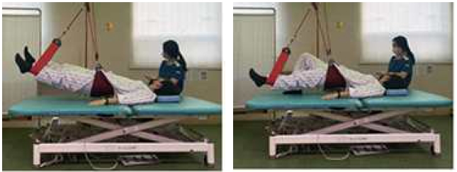
Real-time visual feedback-based sling core stabilization training
(2) Sling core stabilization training
Sling core stabilization training was performed for 30 min, 3 times a week for 4 weeks. The patient performed bridge exercises using a sling device while lying down on a height-adjustable Bobath table. The intensity of exercise was adjusted each week. In the first and second weeks of training, three sets of five repetitions were provided, with 60s of rest between the sets. In the third week of training, the elastic intensity was increased using the red elastic cord, and three sets of five repetitions with 60s of rest between sets were provided. In the fourth week of training, the lower elastic was provided using a black elastic cord, three sets of five repetitions, with a rest period of 60s between the set(Lee et al., 2014) (Table 1).

Sling core stabilization training
(3) General physical therapy
General physical therapy is a one-on-one treatment provided by a physical therapist. Neurologic facilitation approaches such as Bobath's neurodevelopmental therapy and proprioceptive neuromuscular facilitation (PNF) were performed 30 min per session, five times a week, for four weeks.
The trunk impairment scale (TIS), functional reach test (FRT), and postural assessment scale for stroke (PASS) were used to assess dynamic balance. All the evaluations were conducted by a physical therapist with over 10 years of clinical experience and did not treat the participants. Measurements were repeated three times, two days before and two days after the training by the same examiner to minimize measurement errors in an independent laboratory. The average values were used.
In TIS, patients were instructed not to lean on the edge of the mat or treatment table or support themselves with their hands. Further, the patients were instructed to lie on the floor with thighs in complete contact with the mat or table and their feet being as wide apart as the hips. The test commenced with knees bent at 90 degrees and arms resting on the legs. The TIS comprises of three items and the score ranges from a minimum score of 0 to a maximum of 23 points, consisting of static sitting posture balance ability (7 points) and evaluating whether an individual can maintain balance crossing the non-affected lower extremity over the paralyzed lower extremity in a sitting position. Dynamic sitting balance ability evaluates the movement of separating the upper and lower parts of the trunk through lateral flexion in a sitting position (14 points). Coordination ability (6 points), evaluates the horizontal rotational movement of the shoulder and pelvic girdle in a sitting position. Higher scores indicate better trunk control ability (Ko et al., 2015). TIS was found to have a high intraclass correlation coefficient (ICC = 0.96) for test-retest reliability (Verheyden et al., 2004). To measure static and dynamic sitting balance, reliability, efficacy, and internal consistency (Cronbach’s α = 0.89) were reported for patients with stroke (Verheyden et al., 2007).
The functional reach test does not require any equipment and can be easily employed clinically (Wernick-Robinson et al., 1999). It refers to the maximum distance that can be reached forward beyond the length of their arm by extending the arm while maintaining a standing position without moving their feet. This balance test has been widely used to predict falls due to its high reliability and validity(Duncan et al., 1990). It has been reported the arm reach increases with an increase in the size of the trunk and arm movements, and that the trunk plays a crucial role in overall movement (Seidler et al., 2000).
The PASS was used to evaluate postural control performance in patients with stroke, developed by modifying and supplementing the Fugl-Meyer Assessment – Balance (FM-B)(Benaim et al., 2000). Moreover, it is a useful clinical tool for diagnosing the condition of patients with stroke because it can be evaluated easily and within a short duration of 1-10 min (Mao et al., 2002). PASS comprises of three basic postures: lying, sitting, and standing. It consists of 12 items, including five posture maintenance items and seven posture change items. Dynamic balance ability was evaluated as good. PASS has reportedly shown high reliability and validity in patients with stroke (ICC = 0.97; inter-rater reliability, r = 0.98) (Liaw et al., 2012).
Spatiotemporal variables were evaluated using a timed up-and-go (TUG) test and GaitRite to examine walking ability. TUG test assesses functional mobility and dynamic balance. It was used to predict the risk of falls by evaluating the balance ability and functional movement in older individuals. Recently, this test has been used not only for older individuals at high risk of falls, but also for patients with conditions such as stroke, Parkinson’s disease, and arthritis. It can act as a factor in determining the patient’s walking speed, falls, and community walking ability (An et al., 2017). The time taken to return to the chair by going back and forth 3 m after standing up at the command “start” was measured. The patients could use the shoes and aid that they usually wore during the measurement but without the help of the therapist. In this method, the intra-rater reliability was r = 0.99, and the inter-rater reliability was r = 0.98, which was high (Hughes et al., 1998). It is highly valid for evaluating balance, walking speed, and functional movements (Podsiadlo et al., 1991).
The gait ability test measures temporal and spatial gait ability using a gait analyzer (GaitRite, CIR System Inc., USA, 2008) to collect data for quantitative gait analysis of the patient’s gait type. The gait analyzer (GaitRite) is an electronic gait board measuring 5 m in length, 0.6 cm in height, and 61 cm in width. It consists of 16,128 sensors, each with a diameter of 1 cm that are vertically arranged at intervals of 1.27 cm along the walking board to collect information on temporal and spatial variables. Information on the collected temporal and spatial variables was processed using GaitRite GOLD, Version 3.2b (CIR System Inc., USA, 2007) software. The experimental method involved the participant standing in front of a walking board, and when the examiner sent a verbal signal, they walked at the most comfortable speed to move out of the gait board. Gait speed, cadence, step length, stride length, and spatial gait characteristics of single limb support percentage were recorded through informational analysis. The participants were given a 10-minute break between measurements to minimize fatigue. The reliability of this test was r = 0.90 (Van et al., 2004).
3. Data Analysis
All tasks and statistical analyses were performed using SPSS ver. 22.0 (IBM, Chicago, IL, USA). Data were presented as mean and standard deviation. All participants were subjected to Kolmogorov-Smirnov normality verification, confirming that they were normally distributed. Descriptive statistics were used to analyze the general characteristics of the participants. Moreover, homogeneity tests were performed before the experiment by the various variables of the two groups. To determine differences between groups, the experimental results were analyzed using an independent sample t-test. A paired-sample t-test was used to compare before and after training data within the group. The interaction effect between groups over time was analyzed using a two-way repeated measures analysis of variance (ANOVA). The level of statistical significance was set at 0.05.
Ⅲ. Results
1. General and Clinical Characteristics of the Study Participants
A total of 38 post-stroke patients were included in this study. The general and clinical characteristics along with the results of homogeneity tests of the participants are presented in Table 2.
General and clinical characteristics of the study participants(N = 38)
2. Changes in dynamic balance
The group that underwent real-time feedback-based core stabilization training using a sling demonstrated a significant improvement in TIS, FRT, and PASS compared to the control group after 4 weeks of training (p < 0.05). The results revealed a significant interaction between Group × Time, with F-values of F(1, 36) = 36.068, p < 0.001 for TIS; F(1, 36) = 63.890, p < 0.001 for FRT; and F(1, 36) = 89.283, p < 0.001 for PASS (as shown in Table 3). When examining the group effect, only PASS showed a significant difference (p < 0.05).
Changes in dynamic balance and interaction effect(N = 38)
3. Changes in gait parameters
The temporal gait parameters showed a significant interaction between Group × Time effect, with F values of F(1, 36) = 150.893, p < 0.001 in TUG; F(1, 36) = 27.275, p < 0.001 in gait velocity; and F(1, 36) = 54.401, p < 0.001 in cadence. However, no significant group effects were observed(Table 4). The result of spatial gait parameters showed a significant interaction between Group × Time effect with Fvalues of F(1, 36) = 5.601, p = 0.023 in step length of affected side; F(1, 36) = 22.559, p < 0.001 in stride length of affected side; F(1, 36) = 15.516, p < 0.001 in swing phase rate; F(1, 36) = 28.451, p < 0.001 in stance phase rate; F(1, 36) = 34.221, p < 0.001 in single support rate; and F(1, 36) = 22.118, p <0.001 in single support rate. The RFCST group demonstrated a significant difference in terms of all spatial gait parameters in comparison to the control group (p < 0.05)(Table5).
Changes in temporal walking capacity according to the experimental methods(N = 38)
Change in spatial gait parameters according to the experimental method(N = 38)
IV. Discussion
Trunk control and dynamic balance ability are important for independent living and performing activities of daily living (ADL), such as combing, dressing, or going to the bathroom (Ryerson et al., 2008; Isho et al., 2013). Achieving balance in a sitting position after a stroke is the most important factor in rehabilitation, such as maintaining the center of the trunk through adjustments in the lower trunk, pelvis, and lower extremities. This is the first goal to be achieved when treatment is being administered for neurological recovery. The second goal is to minimize activity limitations and improve walking ability to improve the quality of life (Lobo et al., 2013). This study aimed to explore the effects of a four-week intervention involving real-time visual feedback-based sling core stabilization training on dynamic balance and walking ability of 38 patients with stroke.
The changes in dynamic balance before and after training were measured with TIS, FRT, and PASS. The RFCST group showed a significant increase of 1.68 points from 16.95 to 18.63 for TIS, a substantial increase of 3.03 cm from 18.00 cm to 21.03 cm after the experiment in the FRT. In addition, in the PASS test, the RFCST group showed a significant increase of 3.31 points - from 27.32 points before the experiment to 30.63 points after the experiment (p < 0.05). All dynamic balance variables have revealed a considerable time and group interaction effect. However, the minimal clinically important difference (MCID) for TIS reported 3 points in stroke. Our study has achieved only 1.68 points. In a study of 32 patients with stroke, the experimental group that applied core stabilization training for 1 hour, five times a week for four weeks, reported a significant increase in TIS by 2.94 points compared to the control group (p < 0.01), which provides evidence that interventions targeting trunk muscle activation based on core stability theory have positive effects on patients with stroke (Haruyama et al., 2017).
A previous study wherein additional core stabilization training was performed in 1h sessions over five weeks for 110 patients with stroke in the experimental group and 110 individuals in the control group reported improvement in scores of PASS, TIS, Berg Balance Scale (BBS), and Barthel Index (BI) in the experimental group (Cabanas et al., 2021). These results were in concordance with our findings. In a study of 13 patients with stroke, biofeedback treatment was performed using a wearable device that affected motor learning and patient participation and reported an increase in postural maintenance required for dynamic balance and walking (Bowman et al., 2021). In addition, as a result of applying balance training using real-time feedback to 15 patients with stroke for 20 min, three times a week, for four weeks, an increase in BBS balance score was reported (Lupo et al., 2018).
According to a meta-analysis of the effect of SET on balance in patients with stroke, SET treatment combined with conventional rehabilitation was found to be superior than the conventional rehabilitation treatments, with increased degrees of BBS, BI, and Fugl-Meyer Assessment (FMA) in improving post-stroke balance function (Lupo et al., 2018). Patients with neurological disorders, such as stroke, have mobility impairments, including balance and gait disturbances, which increase the risk of falls and affect their quality of life (Jonsdottir et al., 2018). Patients diagnosed with hemiplegia have asymmetric movements and reduced weight-bearing ability due to paralyzed limbs, which in turn affects balance and gait (Wassink et al., 2015). Therefore, the ultimate goal is to improve the weight-bearing ability of paralyzed lower limbs in patients with stroke. Among various treatment methods, training using biofeedback is important to create normal movement patterns (Tate et al., 2010). In this study, core stabilization training using real-time feedback and a sling improved dynamic balance ability, which was consistent with a previous study that reported that imagining movements through visual and auditory stimuli affected weight shift and symmetry of patients with stroke by activating the motor area (Liu et al., 2004). Motion observation, which promotes the reconstruction of brain regions for motion, commands contributes to the formation of observed motor memory and promotes motor memory formation when combined with physical movement (Webster et al., 2006). It has been suggested that motion observation for patients with stroke is a more useful intervention method to improve motor function than simple task-oriented training (Buccino et al., 2006). In this study, it was contemplated that training to control the trunk while providing visual and auditory feedback through the laser beam and verbal instructions of the therapist to help the patients concentrate had a positive effect on the patient’s motor memory.
In addition, core exercises using a sling have been reported to improve trunk stability and increase core muscle strength in a balanced manner, rotate the spine and vertebrae at the apex of left and right curvatures, correct the posture, and maintain the posture to further activate the core muscles (Yu et al., 2019).
The gait pattern of patients with stroke not only shows speed reduction but also confusion regarding weight acceptance and transfer along with inefficient and unstable gait (Nolan et al., 2011). Moreover, movement reportedly decreases in both temporal and spatial gait abilities (Hesse et al., 2008). In this study, changes in gait before and after training were investigated using the TUG test and a spatiotemporal variable evaluation using GaitRite of walking ability. In the TUG test, the RFCST group showed a significant reduction by 2.69 s from 22.70 s before the experiment to 20.01 s after the experiment, and the difference between the groups was found to be statistically significant (p < 0.05). Among the gait variables, the walking speed of the RFCST group increased significantly by 5.57 cm/s, from 37.05 cm/s before the experiment to 42.62 cm/s after the experiment. Moreover, the number of steps increased from 60.76 steps/min before the experiment to 64.68 steps/min after the experiment, demonstrating a significant increase of 3.92 steps /min. This difference between the groups was statistically significant (p < 0.05).
In a study where balance and gait training were conducted using visual feedback for 24 patients with stroke for 30 min, three times a week for eight weeks, the experimental group showed improvements in TUG, BBS evaluation, gait speed, and walking distance compared to the control group (Byl et al., 2015). Moreover, in another study in which weight transfer and gait training was conducted using auditory feedback for 50 minutes, three times a week for six weeks in 24 patients with stroke, the experimental group showed improvement compared to the control group in terms of TUG and 10-meter walk test (10 MWT) evaluations (Choi et al., 2019). In this study, among the gait variables, the affected limb length significantly increased by 3.14 cm from 30.52 cm before the experiment to 33.66 cm after the experiment in the RFCST group, and the difference between the groups was found to be statistically significant (p < 0.05).
In a previous study that showed similar results to ours, recumbent stepping, which requires trunk control, was performed with visual feedback by 11 patients with stroke over six months for 45 min at a time, three times a week for four weeks. The results confirmed that gait-related parameters, including in 5 Times Sit to Stand (5TSTS), Balance Master direction control and speed, and FUGL-Meyer lower extremity function. In the spatiotemporal gait parameter analysis using GaitRite, a significant increase in affected limb guarantee, affected lateral stride, and gait speed has been reported in the experimental group (p < 0.05) (Pardo et al., 2020). In addition, in a study in which core stabilization exercise was performed with real-time feedback training for 30 min a day for six weeks, the experimental group that performed core stabilization exercise through real-time feedback was significantly faster in the TUG test than the control group (p = 0.042). Moreover, stride length (p = 0.021) and single support time (p = 0.033) showed significantly greater improvements in the experimental group than in the control group (Chung et al., 2014).
Visual feedback applied in real-time reportedly affects gait by indicating the degree of motion of the participant at the same time as the motion, which aids in finding motion errors and aptly adjusting them (Tate et al., 2010). Moreover, it is also contemplated that mirror-like visual information is effective for motor learning as it conveys the participant's previous and current movement information, thereby enabling them to realize errors and correct them (Ikegami et al., 2012). Motor learning is noticed to proceed through repeated corrections of motor commands based on motor errors (Shadmehr et al., 2010). In this study, although the concentration and memory of patients with stroke deteriorated, the movement that the participant had to perform was provided with a laser beam to induce interest, and a sling was used to assist weight and focus on the trunk shape, while gravity was removed. This appeared to have increased the concentration.
To improve the ability to walk independently, it is important to promote cognitive functions that govern the walking process. The central nervous system is trained based on the motor learning theory of plasticity of movement, concentration of attention, and repetition of desired movements. Therefore, proprioceptive feedback is necessary for the efficient functioning of the central nervous system to restore and improve walking ability (Pardo et al., 2020). Gait training provides real-time feedback on vertical toe displacement, a gait parameter allowing patients to adjust their toe spacing while walking on a treadmill, which reduces the probability of falling while the patient controls or changes the trajectory of their feet. Gait ability can also be improved. Visual stimulation allows more focus on movement orientation, and through the visual and auditory information provided by the therapist, the patient can easily adjust and correct the trunk by themselves, thereby improving walking ability (Begg et al., 2014; Oouchida et al., 2004).
A limitation of this study is that the small sample size and results obtained in a limited age range make it difficult to generalize the results to all patients with stroke. Moreover, it was also difficult to subdivide and apply the treatment plan or sequence tailored to each patient's functional level. Depending on the quality of the disability, more or less fixed training is possible, and standardized treatment is impossible. Therefore, continuous research on individualized approaches that consider the patient's disability criteria or conditions, various patterns, along family and social environmental influences in realistic treatment situations is needed. Furthermore, it is necessary to evaluate patients with neurological diseases from various aspects, considering that disorders appear in various areas (language and cognitive function, and emotional state), such as complex syndromes, and are not limited to motor dysfunction (Kim et al., 2013).
Ⅳ. Conclusion
Balance issues commonly occur in patients after a stroke. Based on our findings, real-time feedback-based core stabilization training using a sling can be proposed as an effective treatment method for patients with stroke who have insufficient trunk stability. This treatment method can be actively used in clinical practice and would show good results.
Acknowledgments
This paper was supported by the Sahmyook University Research Fund in 2025 (RI12025031).
References
-
Akuthota V, Ferreiro A, Moore T, Fredericson M. Core stability exercise principles. Current sports medicine reports. 2008;7(1):39-44.
[https://doi.org/10.1097/01.CSMR.0000308663.13278.69]

-
An SH, Park D-S, Lim JY. Discriminative validity of the timed up-and-go test for community ambulation in persons with chronic stroke. Physical therapy rehabilitation science. 2017;6(4):176-81.
[https://doi.org/10.14474/ptrs.2017.6.4.176]
-
Benaim C, Pérennou DA, Villy J, Rousseaux M, Pelissier JY. Validation of a standardized assessment of postural control in stroke patients: the Postural Assessment Scale for Stroke Patients (PASS). Stroke. 1999;30(9):1862-8.
[https://doi.org/10.1161/01.STR.30.9.1862]
-
Begg RK, Tirosh O, Said CM, Sparrow WA, Steinberg N, Levinger P, et al. Gait training with real-time augmented toe-ground clearance information decreases tripping risk in older adults and a person with chronic stroke. Frontiers in human neuroscience. 2014;8:243.
[https://doi.org/10.3389/fnhum.2014.00243]
-
Bowman T, Gervasoni E, Arienti C, Lazzarini SG, Negrini S, Crea S, et al. Wearable devices for biofeedback rehabilitation: a systematic review and meta-analysis to design application rules and estimate the effectiveness on balance and gait outcomes in neurological diseases. Sensors. 2021;21(10):3444.
[https://doi.org/10.3390/s21103444]
-
Byl N, Zhang W, Coo S, Tomizuka M. Clinical impact of gait training enhanced with visual kinematic biofeedback: Patients with Parkinson’s disease and patients stable post-stroke. Neuropsychologia. 2015;79:332-43.
[https://doi.org/10.1016/j.neuropsychologia.2015.04.020]
-
Cabanas-Valdés R, Boix-Sala L, Grau-Pellicer M, Guzmán-Bernal JA, Caballero-Gómez FM, Urrútia G. The effectiveness of additional core stability exercises in improving dynamic sitting balance, gait and functional rehabilitation for subacute stroke patients (core-trial): Study protocol for a randomized controlled trial. International Journal of Environmental Research and Public Health. 2021;18(12):6615.
[https://doi.org/10.3390/ijerph18126615]
-
Campbell FM, Ashburn AM, Pickering RM, Burnett M. Head and pelvic movements during a dynamic reaching task in sitting: implications for physical therapists. Archives of physical medicine and rehabilitation. 2001;82(12):1655-60.
[https://doi.org/10.1053/apmr.2001.26818]
-
Chaudhary N, Zafar M, Tariq K, Azam A, Muneer R, Zafar H. Comparative Effectivness of Visual Feedback and Verbal Augmented Feedback for Improving Balance in Stroke Patients. Pakistan Journal of Medical & Health Sciences. 2022;16(12):640-.
[https://doi.org/10.53350/pjmhs20221612640]
-
Chen L, Chen J, Peng Q, Chen J, Zou Y, Liu G. Effect of sling exercise training on balance in patients with stroke: a meta-analysis. PloS one. 2016;11(10):e0163351.
[https://doi.org/10.1371/journal.pone.0163351]
-
Choi Y-H, Kim J-D, Lee J-H, Cha Y-J. Walking and balance ability gain from two types of gait intervention in adult patients with chronic hemiplegic stroke: A pilot study. Assistive technology. 2019;31(2):112-5.
[https://doi.org/10.1080/10400435.2017.1387616]
-
Chung BPH. Effectiveness of robotic-assisted gait training in stroke rehabilitation: A retrospective matched control study. Hong Kong Physiotherapy Journal. 2017;36:10-6.
[https://doi.org/10.1016/j.hkpj.2016.09.001]
-
Chung E, Lee B-H, Hwang S. Core stabilization exercise with real-time feedback for chronic hemiparetic stroke: a pilot randomized controlled trials. Restorative neurology and neuroscience. 2014;32(2):313-21.
[https://doi.org/10.3233/RNN-130353]
-
Clark RA, Pua Y-H, Bryant AL, Hunt MA. Validity of the Microsoft Kinect for providing lateral trunk lean feedback during gait retraining. Gait & posture. 2013;38(4):1064-6.
[https://doi.org/10.1016/j.gaitpost.2013.03.029]
-
Dickstein R, Shefi S, Marcovitz E, Villa Y. Anticipatory postural adjustment in selected trunk muscles in poststroke hemiparetic patients. Archives of physical medicine and rehabilitation. 2004;85(2):261-7.
[https://doi.org/10.1016/j.apmr.2003.05.011]
-
Dean CM, Channon EF, Hall JM. Sitting training early after stroke improves sitting ability and quality and carries over to standing up but not to walking: a randomised controlled trial. Australian Journal of Physiotherapy. 2007;53(2):97-102.
[https://doi.org/10.1016/S0004-9514(07)70042-9]
-
Duncan PW, Weiner DK, Chandler J, Studenski S. Functional reach: a new clinical measure of balance. Journal of gerontology. 1990;45(6):M192-M7.
[https://doi.org/10.1093/geronj/45.6.M192]
-
Giggins OM, Persson UM, Caulfield B. Biofeedback in rehabilitation. Journal of neuroengineering and rehabilitation. 2013;10:1-11.
[https://doi.org/10.1186/1743-0003-10-60]
-
Haruyama K, Kawakami M, Otsuka T. Effect of core stability training on trunk function, standing balance, and mobility in stroke patients: a randomized controlled trial. Neurorehabilitation and neural repair. 2017;31(3):240-9.
[https://doi.org/10.1177/1545968316675431]
-
Hesse S. Treadmill training with partial body weight support after stroke: a review. NeuroRehabilitation. 2008;23(1):55-65.
[https://doi.org/10.3233/NRE-2008-23106]
- Hughes C. Relationship among performance on stair ambulation, functional reach, and timed up and go tests in older adults. Issues Aging. 1998;21:18-22.
-
Ikegami T, Hirashima M, Osu R, Nozaki D. Intermittent visual feedback can boost motor learning of rhythmic movements: evidence for error feedback beyond cycles. Journal of Neuroscience. 2012;32(2):653-7.
[https://doi.org/10.1523/JNEUROSCI.4230-11.2012]
-
Inoue M, Amimoto K, Shida K, Sekine D, Hasegawa D, Fukata K, et al. Effects of Dynamic Sitting Exercise with Delayed Visual Feedback in the Early Post-Stroke Phase: A Pilot Double-Blinded Randomized Controlled Trial. Brain sciences. 2022;12(5):670.
[https://doi.org/10.3390/brainsci12050670]
-
Isho T, Usuda S. Association of trunk control with mobility performance and accelerometry-based gait characteristics in hemiparetic patients with subacute stroke. Gait & posture. 2016;44:89-93.
[https://doi.org/10.1016/j.gaitpost.2015.11.011]
-
Jonsdottir J, Gervasoni E, Bowman T, Bertoni R, Tavazzi E, Rovaris M, et al. Intensive multimodal training to improve gait resistance, mobility, balance, and cognitive function in persons with multiple sclerosis: a pilot randomized controlled trial. Frontiers in neurology. 2018;9:800.
[https://doi.org/10.3389/fneur.2018.00800]
-
Kim S-h, Kwon O-y, Kim S-j, Park K-n, Choung S-d, Weon J-h. Serratus anterior muscle activation during knee push-up plus exercise performed on static stable, static unstable, and oscillating unstable surfaces in healthy subjects. Physical Therapy in Sport. 2014;15(1):20-5.
[https://doi.org/10.1016/j.ptsp.2013.01.001]
-
Kim K-I, Im S-C, Kim K. Effects of trunk stabilization exercises using laser pointer visual feedback in patients with chronic stroke: A randomized controlled study. Technology and Health Care. 2023(Preprint):1-13.
[https://doi.org/10.3233/THC-220100]
- Kim C-R, Min W-K. The effects of virtual reality-based physical therapy in stroke patients. Physical therapy rehabilitation science. 2013;2(1):7-11.
- Kirkesola G. Neurac-a new treatment method for long-term musculoskeletal pain. J Fysioterapeuten. 2009;76(12):16-25.
-
Ko J, You Y. Reliability and responsiveness of the Korean version of the trunk impairment scale for stroke patients. Journal of Korean Physical Therapy. 2015;27(4):175-82.
[https://doi.org/10.18857/jkpt.2015.27.4.175]
-
Langhorne P, Bernhardt J, Kwakkel G. Stroke rehabilitation. The Lancet. 2011;377(9778):1693-702.
[https://doi.org/10.1016/S0140-6736(11)60325-5]
-
Lee JS, Lee HG. Effects of sling exercise therapy on trunk muscle activation and balance in chronic hemiplegic patients. Journal of physical therapy science. 2014;26(5):655-9.
[https://doi.org/10.1589/jpts.26.655]
-
Liaw L-J, Hsieh C-L, Hsu M-J, Chen H-M, Lin J-H, Lo S-K. Test–retest reproducibility of two short-form balance measures used in individuals with stroke. International Journal of Rehabilitation Research. 2012;35(3):256-62.
[https://doi.org/10.1097/MRR.0b013e3283544d20]
-
Liu KP, Chan CC, Lee TM, Hui-Chan CW. Mental imagery for promoting relearning for people after stroke: a randomized controlled trial. Archives of physical medicine and rehabilitation. 2004;85(9):1403-8.
[https://doi.org/10.1016/j.apmr.2003.12.035]
-
Lobo MA, Harbourne RT, Dusing SC, McCoy SW. Grounding early intervention: physical therapy cannot just be about motor skills anymore. Physical therapy. 2013;93(1):94-103.
[https://doi.org/10.2522/ptj.20120158]
- Lupo A, Giovanni Morone M, Cinnera AM, Pucello A, Coiro P, Personeni S, et al. Effects on balance skills and patient compliance of biofeedback training with inertial measurement units and exergaming in subacute stroke: a pilot randomized controlled trial. Functional neurology. 2018;33(3):131-6.
-
Mao H-F, Hsueh I-P, Tang P-F, Sheu C-F, Hsieh C-L. Analysis and comparison of the psychometric properties of three balance measures for stroke patients. Stroke. 2002;33(4):1022-7.
[https://doi.org/10.1161/01.STR.0000012516.63191.C5]
-
Murphy SJ, Werring DJ. Stroke: causes and clinical features. Medicine. 2020;48(9):561-6.
[https://doi.org/10.1016/j.mpmed.2020.06.002]
-
Nolan KJ, Yarossi M. Preservation of the first rocker is related to increases in gait speed in individuals with hemiplegia and AFO. Clinical Biomechanics. 2011;26(6):655-60.
[https://doi.org/10.1016/j.clinbiomech.2011.03.011]
-
Oouchida Y, Okada T, Nakashima T, Matsumura M, Sadato N, Naito E. Your hand movements in my somatosensory cortex: a visuo-kinesthetic function in human area 2. Neuroreport. 2004;15(13):2019-23.
[https://doi.org/10.1097/00001756-200409150-00005]
-
Patterson KK, Gage WH, Brooks D, Black SE, McIlroy WE. Evaluation of gait symmetry after stroke: a comparison of current methods and recommendations for standardization. Gait & posture. 2010;31(2):241-6.
[https://doi.org/10.1016/j.gaitpost.2009.10.014]
-
Pardo V, Albertson M, Bacus M, Crosbie L, Sharkey K, Maher S. Effects of Visual Feedback During Recumbent Stepping in Individuals With Chronic Stroke. Archives of rehabilitation research and clinical translation. 2020;2(4):100086.
[https://doi.org/10.1016/j.arrct.2020.100086]
-
Podsiadlo D, Richardson S. The timed “Up & Go”: a test of basic functional mobility for frail elderly persons. Journal of the American Geriatrics Society. 1991;39(2):142-8.
[https://doi.org/10.1111/j.1532-5415.1991.tb01616.x]
-
Quaney BM, He J, Timberlake G, Dodd K, Carr C. Visuomotor training improves stroke-related ipsilesional upper extremity impairments. Neurorehabilitation and neural repair. 2010;24(1):52-61.
[https://doi.org/10.1177/1545968309341646]
-
Ryerson S, Byl NN, Brown DA, Wong RA, Hidler JM. Altered trunk position sense and its relation to balance functions in people post-stroke. Journal of Neurologic Physical Therapy. 2008;32(1):14-20.
[https://doi.org/10.1097/NPT.0b013e3181660f0c]
- Schmidt RA, Lee TD, Winstein C, Wulf G, Zelaznik HN. Motor control and learning: A behavioral emphasis: Human kinetics; 2018.
-
Seidler RD, Stelmach GE. Trunk-assisted prehension: specification of body segments with imposed temporal constraints. Journal of motor behavior. 2000;32(4):379-89.
[https://doi.org/10.1080/00222890009601387]
-
Shadmehr R, Smith MA, Krakauer JW. Error correction, sensory prediction, and adaptation in motor control. Annual review of neuroscience. 2010;33:89-108.
[https://doi.org/10.1146/annurev-neuro-060909-153135]
-
Sun X, Gao Q, Dou H, Tang S. Which is better in the rehabilitation of stroke patients, core stability exercises or conventional exercises? Journal of physical therapy science. 2016;28(4):1131-3.
[https://doi.org/10.1589/jpts.28.1131]
-
Tate JJ, Milner CE. Real-time kinematic, temporospatial, and kinetic biofeedback during gait retraining in patients: a systematic review. Physical therapy. 2010;90(8):1123-34.
[https://doi.org/10.2522/ptj.20080281]
- Vaishnavi W, Ragini D, Mangulkar UR. Use of Robotics in Gait Rehabilitation Following Stroke: A Review. Cureus. 2022;14(11).
-
Van Criekinge T, Saeys W, Hallemans A, Vereeck L, De Hertogh W, Van de Walle P, et al. Effectiveness of additional trunk exercises on gait performance: study protocol for a randomized controlled trial. Trials. 2017;18:1-12.
[https://doi.org/10.1186/s13063-017-1989-1]
-
Van Uden CJ, Besser MP. Test-retest reliability of temporal and spatial gait characteristics measured with an instrumented walkway system (GAITRite®). BMC Musculoskeletal Disorders. 2004;5:1-4.
[https://doi.org/10.1186/1471-2474-5-13]
-
Verheyden G, Nieuwboer A, Mertin J, Preger R, Kiekens C, De Weerdt W. The Trunk Impairment Scale: a new tool to measure motor impairment of the trunk after stroke. Clinical rehabilitation. 2004;18(3):326-34.
[https://doi.org/10.1191/0269215504cr733oa]
-
Verheyden G, Nieuwboer A, De Wit L, Feys H, Schuback B, Baert I, et al. Trunk performance after stroke: an eye catching predictor of functional outcome. Journal of neurology, neurosurgery & psychiatry. 2007;78(7):694-8.
[https://doi.org/10.1136/jnnp.2006.101642]
-
Wassink G, Barrett RD, Davidson JO, Bennet L, Galinsky R, Dragunow M, et al. Hypothermic neuroprotection is associated with recovery of spectral edge frequency after asphyxia in preterm fetal sheep. Stroke. 2015;46(2):585-7.
[https://doi.org/10.1161/STROKEAHA.114.008484]
-
Wernick-Robinson M, Krebs DE, Giorgetti MM. Functional reach: does it really measure dynamic balance? Archives of physical medicine and rehabilitation. 1999;80(3):262-9.
[https://doi.org/10.1016/S0003-9993(99)90136-3]
-
Webster BR, Celnik PA, Cohen LG. Noninvasive brain stimulation in stroke rehabilitation. NeuroRx. 2006;3(4):474-81.
[https://doi.org/10.1016/j.nurx.2006.07.008]
-
Buccino G, Solodkin A, Small SL. Functions of the mirror neuron system: implications for neurorehabilitation. Cognitive and behavioral neurology. 2006;19(1):55-63.
[https://doi.org/10.1097/00146965-200603000-00007]
-
Yoon S-W, Son H-H. Effect of Visual Feedback Training of Core Strength on Coordination, Balance and Walking Ability of Stroke Patients. Korean Society of Physical Medicine. 2020;15(4):145-53.
[https://doi.org/10.13066/kspm.2020.15.4.145]
- Yu D-y, Yang Y-s, Park S-d. Effects of Sling-Based Core Exercise Program on Cobb Angle and Core Strength of Idiopathic Scoliosis in Adolescences. The Journal of Korean Academy of Orthopedic Manual Physical Therapy. 2019;25(2):47-56.
-
Yoo G-t. Effects of Core Stabilization Exercise According to Mirror Visual Feedback and Video Visual Feedback on Static and Dynamic Balance in College students with Lumbar Back Pain. The Journal of Korean Academy of Physical Therapy Science. 2025;32(4):95-110
[https://doi.org/10.26862/jkpts.2025.12.32.4.95]
-
Kim J-h, Park S-b, Im W-s. Comparison of the Immediate Effects of Different Intervention Times in a Visual Feedback-based Balance Training Program. The Journal of Korean Academy of Physical Therapy Science. 2025;32(1):19-27
[https://doi.org/10.26862/jkpts.2025.03.32.1.19]