
Effects of Modified Environmental and Task Conditions of the Single-Leg Bridge on Lower Limb Muscle Activity in Healthy Adults: A Cross-Sectional Study
© 2026 by the Korean Physical Therapy Science
Abstract
This study was to investigate the effects of visual deprivation and unstable surface conditions on lower-limb muscle activation during the single leg bridge exercise. Supporting limb strength is essential for maintaining balance and functional movement, yet how sensory and environmental factors modify muscle recruitment during this exercise remains unclear.
Cross-sectional Study
Forty-two healthy adults aged 30–40 years participated. They performed four types of single leg bridge exercises: basic (SB), with visual deprivation (SBV), on an unstable surface (SBU), and on an unstable surface with visual deprivation (SBUV). Activation of the rectus femoris, vastus medialis oblique, vastus lateralis, tibialis anterior, gastrocnemius, biceps femoris, and gluteus maximus was measured using surface electromyography, and EMG signals were normalized to each participant’s maximum voluntary isometric contraction.
The results showed that muscle activation increased progressively in SBV, SBU, and SBUV conditions compared to SB. The gluteus maximus, hamstrings, and gastrocnemius showed the greatest increases, with the highest overall activation occurring in the SBUV condition.
These results suggest that modifying environmental and sensory conditions during the single leg bridge exercise effectively enhances lower limb muscle activation, particularly in the gluteus maximus and hamstrings. Stepwise application of these variations may serve as an effective strategy for rehabilitation, balance improvement, and fall prevention in both clinical and athletic populations.
Keywords:
Bridge therapy, Electromyography, Lower limb, Muscle strength, Visual deprivationⅠ. Introduction
Lower limb strength is essential for performing daily activities and maintaining independent mobility (Muehlbauer et al., 2015). In particular, basic functional movements such as walking, stair climbing, and rising from a chair presuppose an adequate level of lower limb strength (Atar et al., 2021). Adequate lower limb strength contributes to balance control and postural stability, both of which are important for fall prevention (Clemson et al., 2012). It also supports greater participation in physical activities and is associated with improved functional performance and quality of life. (Khodadad Kashi et al., 2023). Therefore, maintaining lower limb strength is important for healthy aging and functional independence. When lower limb strength is weakened, the risk of falls increases due to reduced walking ability and difficulty in maintaining balance, and the limitation of daily living activities may hinder the maintenance of independent living (Muehlbauer et al., 2015). This, in the long term, leads to complex secondary problems such as muscle atrophy, reduced cardiovascular function, and psychological decline due to decreased physical activity (Hayashi et al., 2021), ultimately severely impairing an individual's quality of life (Fábrega-Cuadroset al., 2021). Reduced lower limb strength may limit physical activity and independence, which can negatively affect both physical and psychological well-being.
Among various interventions for strengthening lower limb muscles, the bridge exercise is known as a relatively safe and easy-to-perform, weight-bearing exercise that can effectively activate the gluteus maximus and hamstring muscles (Kellis et al., 2023). In particular, the bridge exercise performed by lifting the pelvis while lying on the back can be applied across all age groups and has been widely used in rehabilitation settings (Hollman et al., 2018). The single-leg bridge is a variation of the bridge exercise that increases loading on one limb, thereby promoting greater muscle activation (Kim & Park, 2016). It has been recognized as an effective strategy for improving the strength of the non-dominant lower limb and enhancing neuromuscular control (García-Vaqueroet al., 2012). The single leg bridge exercise can provide a meaningful mechanism for addressing strength imbalances between both lower limbs and enhancing proprioceptive stimulation (Choi et al., 2016), and it possesses characteristics that contribute to high-intensity muscle stimulation of the ipsilateral lower limb and improvement of core stability beyond simple repetitive movements (Youdas et al., 2017). Furthermore, due to its ability to be performed without spatial constraints, it offers high accessibility and its applicability is gradually expanding not only for patient populations aiming for functional recovery but also in sports rehabilitation and training programs for high-performance athletes (Prat-Luri et al., 2025). Although several studies have investigated variations of the bridge exercise and their associated electromyographic responses, most have focused on trunk stabilization or hip muscle activation under stable surface conditions. However, studies specifically comparing muscle activation patterns during the single-leg bridge under different environmental and sensory conditions remain limited. The patterns of muscle action and recruitment strategies may vary depending on the exercise environment and loading conditions, which provide essential foundational data for optimizing intervention effects and developing tailored exercise prescriptions. Therefore, this study aimed to investigate the effects of visual deprivation and unstable surface conditions during the single-leg bridge exercise on lower limb muscle activation in healthy adults.
Ⅱ. Methods
1. Participants
This study recruited participants by posting a notice on the bulletin board of C University in Jeollanam-do, targeting healthy adults aged 30–40 years who voluntarily expressed their willingness to participate. The study objectives and procedures were thoroughly explained, and participants were informed that they could withdraw at any time without any disadvantage. The inclusion criteria were as follows: 1) individuals without orthopedic disorders, 2) individuals with joint mobility within the normal range, 3) individuals with no history of surgery, 4) individuals without recent trauma or pain, and 5) individuals not diagnosed with osteoporosis (Cho et al., 2024). The exclusion criteria were as follows: 1) individuals with lumbar radiculopathy or vascular claudication, 2) individuals with neurological disorders or lower-extremity musculoskeletal conditions that could interfere with performing the single-leg bridge exercise, 3) individuals with cardiovascular or respiratory diseases that limit moderate physical activity, and 4) individuals who had experienced lower-extremity pain, surgery, trauma, or rehabilitation treatment within the past six months(Amundsen et al., 2018; Kilpikoski et al., 2024). The required sample size was calculated using G*Power 3.1.9.7 (Faul et al., 2007). Based on a repeated-measures one-way ANOVA with an effect size of 0.5, a power of 0.95, and a significance level of 0.05, a minimum of 24 participants was determined to be necessary (Kim et al., 2023).Considering potential dropouts, a total of 50 participants were recruited, and ultimately, after excluding 8 participants who withdrew during the study, 42 participants were included in the analysis. This study was approved by the Institutional Review Board of S Rehabilitation Hospital (IRB No. 202509-WR-02).
2. Experimental Procedures
The exercise interventions applied in this study consisted of four types: the single leg bridge (SB), the single leg bridge with visual deprivation (SBV), the single leg bridge on an unstable surface (SBU), and the single leg bridge on an unstable surface with visual deprivation (SBUV). The order of performing each exercise was determined by random draw.
Muscle activation was assessed using surface electromyography (sEMG) of the rectus femoris (RF), vastus medialis oblique (VMO), vastus lateralis (VL), tibialis anterior (TA), gastrocnemius (GCM), biceps femoris (BF), and gluteus maximus (GM). Electrodes were attached following SENIAM guidelines, parallel to the direction of the muscle fibers (Hermens et al., 2000).
EMG signals were collected at 1500 Hz and band-pass filtered between 20 and 500 Hz. Signals were full-wave rectified, and RMS values were calculated at 500 ms intervals. A 3-second segment excluding the first and last 1 second of each measurement period was selected for analysis. Measurements were repeated three times, and mean values were used. Muscle activity was normalized to maximum voluntary isometric contraction (MVIC) and expressed as a percentage (Kim et al., 2023). Body hair was removed, and skin was disinfected before electrode attachment (Kusche et al., 2018). During MVIC, an assistant researcher stabilized the lower limbs to minimize movement (Buckthorpe et al., 2012).
Prior to the experiment, participants practiced each posture at least three times. Maximum muscle activity was measured for 5 seconds for each muscle (RF, VMO, VL, TA, GCM, BF, GM). The four exercises were performed as follows:
SB: Participants performed a single leg bridge using the supporting limb on a Bobath table. The hip and knee joints were maintained at 90° flexion, the lumbar region in neutral alignment, and the upper limbs relaxed. The pelvis and trunk were raised approximately 10cm to selectively activate the supporting limb muscles, while the researcher monitored and provided guidance to minimize compensatory movements <Fig. 1>.
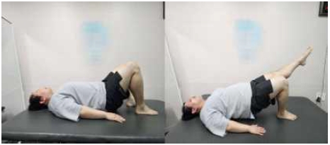
Single Leg Bridge
SBV: Participants performed a single leg bridge on a Bobath table while wearing a blindfold. The hip and knee joints were maintained at 90° flexion, the lumbar region in neutral alignment, and the upper limbs relaxed. The supporting limb foot remained on the ground, and the pelvis and trunk were raised approximately 10cm. The researcher continuously monitored the participants and provided guidance to minimize compensatory movements <Fig. 2>.
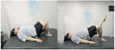
Single leg Bridge with Visual deprivation
SBU: Participants performed a single leg bridge on an unstable surface by placing a balance pad under the supporting limb foot. The hip and knee joints were maintained at 90° flexion, the lumbar region in neutral alignment, and the upper limbs relaxed. The pelvis and trunk were raised approximately 10cm, and the researcher provided repeated feedback to minimize compensatory movements <Fig. 3>.
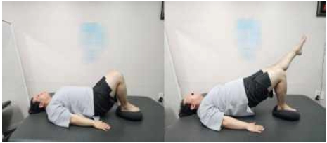
Single leg Bridge on an Unstable surface
SBUV: Participants performed a single leg bridge on a Bobath table with the supporting limb foot fixed. The hip and knee joints were maintained at 90° flexion, the lumbar region in neutral alignment, and the upper limbs relaxed. The pelvis and trunk were raised approximately 10cm, and the researcher provided verbal and tactile feedback to ensure proper posture and minimize compensatory movements <Fig. 4>.
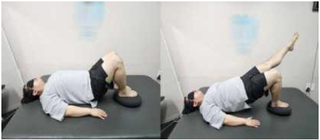
SBUV: Single leg Bridge on an Unstable surface with Visual deprivation
3. Statistical Analysis
Statistical analyses and data processing for this study were performed using SPSS Statistics version 28.0 (IBM Corp., Armonk, NY, USA), and the mean and standard deviation of each variable were calculated. Normality of the data was assessed using the Shapiro-Wilk test, and participants’ general characteristics were analyzed using descriptive statistics. To compare changes in muscle activity during the single leg bridge exercise across conditions, repeated measures one-way ANOVA was conducted. The least significant difference (LSD) test was used for post hoc comparisons between conditions. The significance level (α) was set at 0.05 for all statistical analyses.
Ⅲ. Results
This study compared lower limb muscle activity under four conditions in 42 healthy adults <Table 1>. Significant differences were observed among conditions for all muscle groups (p<.05). Muscle activity progressively increased in the SBV, SBU, and SBUV conditions compared to SB. The greatest increases were observed in the GM, HS, and GCM. Overall, the highest muscle activation was observed under the SBUV condition, in which both an unstable surface and visual deprivation were applied simultaneously <Table 2>.

General characteristics of the participants
Muscle activation of lower extremity muscles during single leg bridge exercises with different conditions(%)
Ⅳ. Discussion
This study analyzed changes in lower limb muscle activity according to the performance conditions of the single leg bridge exercise, and significant differences were observed among conditions for all muscle groups. Notably, the highest muscle activation was observed when both an unstable surface and visual manipulation were applied, with pronounced increases in the GM, HS, and GCM. These findings suggest that the single leg bridge exercise not only selectively strengthens hip extensor muscles but can also maximize neuromuscular responses through manipulation of the exercise environment.
First, the effects of exercise on an unstable surface can be interpreted as a result of enhanced proprioceptive input and trunk control to maintain balance and postural stability. Previous studies have reported that exercises on unstable surfaces promote neuromuscular coordination and simultaneous activation of muscle fibers, thereby eliciting higher electromyographic responses (Choi et al., 2016). In the present study, muscle activity in all muscle groups was significantly increased under the SBU and SBUV conditions, supporting this mechanism.
Second, the influence of the presence or absence of vision is also noteworthy. The availability of visual input allows participants to monitor their surroundings and maintain trunk and lower limb alignment, which can minimize unnecessary compensatory movements and promote efficient recruitment of primary muscles. In contrast, visual deprivation increases reliance on vestibular and somatosensory inputs due to the lack of visual cues, leading to greater demands for postural stabilization and consequently higher muscle activation. This indicates that the presence or absence of visual information is an important modulating factor for exercise performance strategies and neuromuscular responses (Hollman et al., 2018).
Third, the differences in muscle specific activation are also noteworthy. Notably, pronounced increases were observed in the GM and HS, which can be attributed to the primary movement of the single leg bridge exercise involving hip extension. Previous studies have reported that bridge variations selectively strengthen the GM and HS muscle groups (Kellis et al., 2023; Vincent et al., 2019), and the findings of the present study are consistent with these reports. Furthermore, increased activation of the RF,VMO, VL was also observed, suggesting that the single leg bridge exercise positively influences not only the hip extensors but also muscles involved in hip and knee joint stability.
Fourth, the findings of this study have significant clinical implications. Lower limb strength is essential for performing daily functional activities such as walking, standing, and maintaining balance (Muehlbauer et al., 2015; Clemson et al., 2012), and muscle weakness can increase the risk of falls and reduce functional independence. Therefore, as demonstrated in this study, the single leg bridge exercise with modulated execution conditions can serve as a practical intervention strategy in rehabilitation, allowing stepwise application according to the patient’s characteristics and functional level. In particular, it can be effectively applied in rehabilitation for older adults aimed at improving balance and preventing falls, as well as in sports rehabilitation programs targeting lower limb strength asymmetries.
Finally, it is necessary to acknowledge the limitations of this study. First, the participants were limited to healthy adults aged 30–40 years, which restricts the generalizability of the findings to older adults or clinical populations. Second, this study measured only short-term electromyographic responses, so it could not determine whether long-term exercise would lead to actual improvements in muscle strength or functional performance. Therefore, future research should include long-term follow-up studies encompassing diverse age groups and patient populations. Additionally, it would be desirable to comprehensively examine the relationships between muscle activity and functional outcomes, such as balance assessments and gait analysis.
Ⅴ. Conclusion
This study demonstrated that performing the single leg bridge exercise under altered surface stability and visual conditions significantly increased activation of major lower limb muscles, with the greatest effects observed in the gluteus maximus and hamstrings. These findings suggest that manipulating exercise conditions can maximize neuromuscular stimulation during the single leg bridge. Accordingly, this exercise can be effectively applied in clinical rehabilitation and exercise prescription, with difficulty progressively adjusted according to individual characteristics and capabilities. The single leg bridge thus holds potential as an intervention for enhancing lower limb strength, improving pelvic stability, promoting balance, and preventing falls.
References
-
Amundsen PA, Evans DW, Rajendran D, et al. Inclusion and exclusion criteria used in non-specific low back pain trials: a review of randomised controlled trials published between 2006 and 2012. BMC Musculoskelet Disord. 2018;19:113.
[https://doi.org/10.1186/s12891-018-2034-6]

-
Atar S, Demirhan E, Cabuk H, et al. Comparison of pain, muscle strength, and functional status following unicompartmental knee arthroplasty, total knee arthroplasty, and conservative management of gonarthrosis. Indian J Orthop. 2021 Oct 26;56(3):464-72.
[https://doi.org/10.1007/s43465-021-00549-5]
- Buckthorpe MW, Hannah R, Pain TG, et al. Reliability of neuromuscular measurements during explosive isometric contractions, with special reference to electromyography normalization techniques. Muscle Nerve. 2012 Oct;46(4):566-76.
-
Choi K, Bak J, Cho M, et al. The effects of performing a one-legged bridge with hip abduction and use of a sling on trunk and lower extremity muscle activation in healthy adults. J Phys Ther Sci. 2016 Sep;28(9):2625-8.
[https://doi.org/10.1589/jpts.28.2625]
-
Cho M, Shin H, Chung Y. The effect of dominant side leg exercise in different environments with and without resistance on non-dominant leg muscle activation: sitting position. Physical Therapy Rehabilitation Science. 2024;13(4):534-543.
[https://doi.org/10.14474/ptrs.2024.13.4.534]
-
Clemson L, Fiatarone Singh MA, Bundy A, et al. Integration of balance and strength training into daily life activity to reduce rate of falls in older people (the LiFE study): randomised parallel trial. BMJ. 2012 Aug 7;345:e4547.
[https://doi.org/10.1136/bmj.e4547]
-
Fábrega-Cuadros R, Hita-Contreras F, Martínez-Amat A, et al. Associations between the severity of sarcopenia and health-related quality of life in community-dwelling middle-aged and older adults. Int J Environ Res Public Health. 2021 Jul 29;18(15):8026.
[https://doi.org/10.3390/ijerph18158026]
-
Faul F, Erdfelder E, Lang AG, et al. G* Power 3: A flexible statistical power analysis program for the social, behavioral, and biomedical sciences. Behavior Research Methods. 2007;39(2):175-191.
[https://doi.org/10.3758/BF03193146]
-
García-Vaquero MP, Moreside JM, Brontons-Gil E, et al. Trunk muscle activation during stabilization exercises with single and double leg support. J Electromyogr Kinesiol. 2012 Jun;22(3):398-406.
[https://doi.org/10.1016/j.jelekin.2012.02.017]
-
Hayashi C, Ogata S, Okano T, et al. Long-term participation in community group exercise improves lower extremity muscle strength and delays age-related declines in walking speed and physical function in older adults. Eur Rev Aging Phys Act. 2021 May 28;18(1):6.
[https://doi.org/10.1186/s11556-021-00260-2]
-
Hermens HJ, Freriks B, Disselhorst-Klug C, et al. Development of recommendations for SEMG sensors and sensor placement procedures. J Electromyogr Kinesiol. 2000 Oct;10(5):361-74.
[https://doi.org/10.1016/S1050-6411(00)00027-4]
-
Hollman JH, Berling TA, Crum EO, et al. Do verbal and tactile cueing selectively alter gluteus maximus and hamstring recruitment during a supine bridging exercise in active females? A randomized controlled trial. J Sport Rehabil. 2018 Mar 1;27(2):138-43.
[https://doi.org/10.1123/jsr.2016-0130]
-
Kellis E, Konstantopoulos A, Ellinoudis A. Effect of bridge exercise duration on lateral abdominal muscle thickness and gluteus maximus activation. J Sport Rehabil. 2023 May 24;32(7):773-81.
[https://doi.org/10.1123/jsr.2022-0286]
-
Kilpikoski S, Häkkinen AH, Repo JP, et al. The McKenzie Method versus guideline-based advice in the treatment of sciatica: 24-month outcomes of a randomised clinical trial. Clinical Rehabilitation. 2023;38(1):72-84.
[https://doi.org/10.1177/02692155231196393]
-
Kim H, Cho M, Chung Y. Effects of hip flexion by sitting posture on trunk muscle activity and balance in healthy adults. Phys Ther Rehabil Sci. 2023;12(4):398-403.
[https://doi.org/10.14474/ptrs.2023.12.4.398]
-
Kim J, Park M. Changes in the activity of trunk and hip extensor muscles during bridge exercises with variations in unilateral knee joint angle. J Phys Ther Sci. 2016 Sep;28(9):2537-40.
[https://doi.org/10.1589/jpts.28.2537]
-
Kusche R, Kaufmann S, Ryschka M. Dry electrodes for bioimpedance measurements—Design, characterization and comparison. Biomed Phys Eng Express. 2018;5(1):015001.
[https://doi.org/10.1088/2057-1976/aaea59]
-
Muehlbauer T, Gollhofer A, Granacher U. Associations between measures of balance and lower-extremity muscle strength/power in healthy individuals across the lifespan: a systematic review and meta-analysis. Sports Med. 2015 Dec;45(12):1671-92.
[https://doi.org/10.1007/s40279-015-0390-z]
- Prat-Luri A, Vera-Garcia FJ, Moreno-Navarro P, et al. Short-term bridging and bird-dog exercise programs did not enhance trunk performance and whole-body dynamic balance in young physically active males: a double-blind randomized trial. PLoS One. 2025 Jun 5;20(6):e0325040.
-
Youdas JW, Hartman JP, Murphy BA, et al. Electromyographic analysis of gluteus maximus and hamstring activity during the supine resisted hip extension exercise versus supine unilateral bridge to neutral. Physiother Theory Pract. 2017 Feb;33(2):124-30.
[https://doi.org/10.1080/09593985.2016.1271848]